. 2011 Feb 24;117(8):2484-93.
doi: 10.1182/blood-2010-05-284653. Epub 2010 Dec 16.

Neoangiogenesis contributes to the development of hemophilic synovitis

Affiliations

Neoangiogenesis contributes to the development of hemophilic synovitis

Suchitra S Acharya et al. Blood. .

Abstract

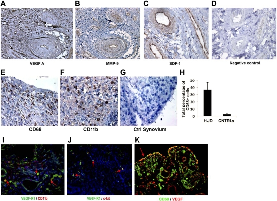
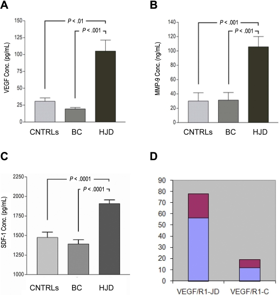
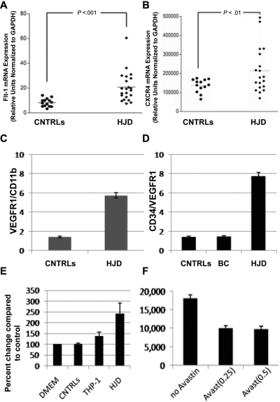
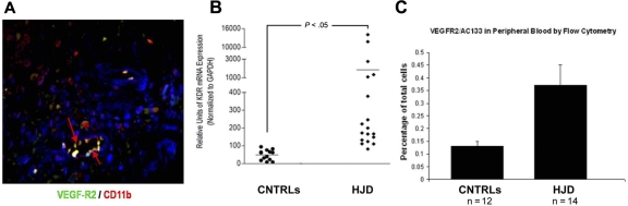
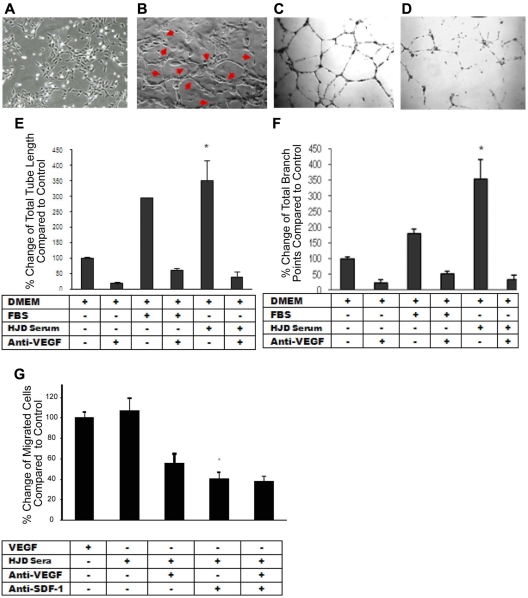
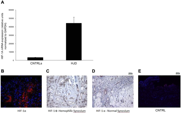
Joint arthropathy secondary to recurrent hemarthroses remains a debilitating complication of hemophilia despite the use of prophylactic factor concentrates. Increased vascularity and neoangiogenesis have been implicated in the progression of musculoskeletal disorders and tumor growth. We hypothesized that de novo blood vessel formation could play a major role in the pathogenesis of hemophilic joint disease (HJD). We observed a 4-fold elevation in proangiogenic factors (vascular endothelial growth factor-A [VEGF-A], stromal cell-derived factor-1, and matrix metalloprotease-9) and proangiogenic macrophage/monocyte cells (VEGF(+)/CD68(+) and VEGFR1(+)/CD11b(+)) in the synovium and peripheral blood of HJD subjects along with significantly increased numbers of VEGFR2(+)/AC133(+) endothelial progenitor cells and CD34(+)/VEGFR1(+) hematopoietic progenitor cells. Sera from HJD subjects induced an angiogenic response in endothelial cells that was abrogated by blocking VEGF, whereas peripheral blood mononuclear cells from HJD subjects stimulated synovial cell proliferation, which was blocked by a humanized anti-VEGF antibody (bevacizumab). Human synovial cells, when incubated with HJD sera, could elicit up-regulation of HIF-1α mRNA with HIF-1α expression in the synovium of HJD subjects, implicating hypoxia in the neoangiogenesis process. Our results provide evidence of local and systemic angiogenic response in hemophilic subjects with recurrent hemarthroses suggesting a potential to develop surrogate biologic markers to identify the onset and progression of hemophilic synovitis.


Figures

Figure 1
Figure 1
Hemophilic synovium expresses proangiogenic mediators and myeloid cells. Immunohistochemical analyses using DAB revealed positive staining for proangiogenic mediators VEGF-A (A), MMP-9 (B), and SDF-1 (C), in hemophilic synovium but not control synovium (D) (original magnification ×200). Immunohistochemical staining for CD68 (E) and CD11b (F) provided evidence for the presence of myeloid cell infiltration not observed in control synovium (G). Quantitative assessment of CD68+ cells in hemophilic and control synovium demonstrated an increased percentage of CD68+ cells in hemophilic synovium (HJD) compared with control (CNTRLs) (H). Immunofluorescent staining (red arrow) demonstrates CD11b+ cells coexpressing VEGF-R1 (I) in hemophilic synovium. Hemophilic synovium also contained VEGFR1 (red arrows) cells coexpressing the early HPC marker c-kit (J). These CD68+ cells expressed VEGF showing coexpression within the hemophilic synovium (K). Total number of sections stained for both experimental and control groups detailed in “HJD synovium expresses proangiogenic mediators and myeloid cells” (original magnification ×400).
Figure 2
Figure 2
Angiogenic mediators are elevated in the plasma of HJD subjects. Enzyme-linked immunosorbent assays were performed as per the manufacturer's protocol using anti VEGF-A, anti-MMP-9, anti-SDF-1 antibodies. (A) A 4-fold elevation of plasma VEGF-A was observed in HJD subjects (prospective cohort) compared with control group A (BC; P < .001) and control group B (CNTRLs; P < .01). (B) MMP-9 levels were also significantly elevated in HJD subjects (prospective cohort) compared with control group A (BC; P < .001) and control group B (CNTRLs; P < .001). (C) SDF-1 levels were also significantly elevated in HJD subjects compared with control group A (BC; P < .0001) and control group B (CNTRLs; P < .0001). (D) These subjects with elevated VEGF levels also had increased expression of VEGFR1 mRNA (VEGF/R1-JD) compared with control groups A and B grouped together (VEGF/R1-C; P < .05). Data are mean ± SEM.
Figure 3
Figure 3
Circulating levels of HPCs expressing VEGFR1 and CXCR4 are increased in peripheral blood of HJD subjects. (A) mRNA from the peripheral blood of HJD subjects (prospective cohort as defined in “Subjects and samples”) show significant up-regulation of VEGFR1 mRNA (labeled Flt-1 mRNA, mouse equivalent of human VEGFR1; P < .001; A) and CXCR4 (P < .01; B) compared with controls (CNTRLs, control group A). Results represent data from 3 experiments. (C) Circulating HPCs (VEGFR1+/CD11b+) are elevated 4-fold in the peripheral blood of HJD subjects compared with controls by flow cytometry. (D) HPCs, which express CD34/VEGFR1-early myeloid cells, were also increased 4-fold in HJD subjects compared with control group A (BC) and control group B (CNTRLs) by flow cytometry. (E) PBMCs from subjects with HJD increased synovial cell proliferation by 2.5-fold compared with medium alone (DMEM) and control groups A and B (CNTRLs). THP-1 cells (immortalized cells of the monocyte/macrophage lineage used as a positive control) stimulated synovial proliferation up to 1.5-fold (THP-1). These results are expressed as a percentage change compared with the controls. (F) Synovial proliferation by PBMCs from HJD subjects (prospective cohort) could be abrogated by Avastin (Avast), an inhibitor of VEGF, by 50% using 2 different concentrations (0.25 mg/mL, 0.5 mg/mL) of the drug. These results are expressed as number of cells before and after treatment with Avastin.
Figure 4
Figure 4
Endothelialization occurs in HJD synovium. (A) HJD synovium coexpressed VEGFR2/CD 11b (indicated by yellow staining with red arrows), late EPC markers. (B) VEGFR2 mRNA (labeled kdr, mouse equivalent of human VEGFR2) levels as quantified by quantitative PCR were increased in the peripheral blood of HJD subjects compared with control groups A and B (CNTRLs). (C) Significantly increased numbers of VEGFR2+/AC133+ early EPCs were also observed in the peripheral blood of HJD subjects compared with control groups A and B (CNTRLs). These data are expressed as a percentage of total peripheral blood mononuclear cells analyzed by flow cytometry.
Figure 5
Figure 5
HJD plasma elicits an angiogenic response from ECs that is VEGF-dependent. (A) Endothelial cells incubated with control group A sera for 22 hours showing normal morphology. (B) Endothelial cells incubated with HJD sera for 22 hours revealing the presence of “tube formation” representing blood vessels as indicated by the red arrows. Results replicated 4 times and represent data from 1 experiment. Human umbilical vein endothelial cells grown in DMEM culture medium (10 000/well) were plated on Matrigel-coated 48-well plates with (D) or without (C) preincubation with anti-VEGF antibody (10 μg/mL) and with or without FBS or HJD sera. Images reflected tube formation after 16-hour incubation detected by phase-contrast microscopy (original magnification ×40). Representative results from 4 experiments. Quantitative assessment of mean and total tube lengths and branch points using Metamorph software Version 40002 (Molecular Devices) revealed an 84% reduction in total tube length (SEM). *P < .001. (E) An 82% reduction in mean (SEM) tube length (data not shown). P < .001. A 93% reduction in mean (SEM) branch points. P < .001. (F) Representative results from 3 experiments. Migration across a transwell barrier was induced by VEGF and HJD serum and abrogated by anti-VEGF and anti–SDF-1 antibody. When human umbilical vein endothelial cells were plated on 24-well plates with or without VEGF, HJD serum, anti-VEGF blocking peptide, or anti-SDF-1 antibody, a 55% reduction in human umbilical vein endothelial cell migration with anti-VEGF antibody and a 60% reduction with anti-SDF-1 antibody (P < .01) was observed. There was no additive effect of anti-VEGF and anti–SDF-1 antibodies. (G) Results are expressed as a percent of the control, which is human umbilical vein endothelial cells grown in the presence of VEGF. Representative results from 3 experiments.
Figure 6
Figure 6
Sera from HJD subjects induce expression of HIF-1α in synovial cells. (A) Synovial cells were incubated with HJD serum and serum from control groups A and B for 10, 30, and 60 minutes, after which mRNA was extracted and subjected to quantitative PCR. (A) Significantly increased mRNA expression for HIF-1α was observed at 60 minutes in cells incubated with HJD serum compared with control serum (CNTRLs; P < .0001). No significant difference was observed at the 10- or 30-minute time points. Results represent data from 3 independent experiments. (B) Immunofluorescence of hemophilic synovium expressing HIF-1α. Immunofluorescence using a monoclononal antibody against HIF-1α demonstrated abundant expression of HIF-1α in hemophilic synovium in the endothelial cells and lining cells (B) but not in control synovium (E). Similarly, immunohistochemical staining using DAB confirmed HIF-1α positivity in similar regions in hemophilic synovium (C, original magnification ×40) and expression of HIF-1α only in endothelial cells in normal synovium (D, original magnification ×40). Results were replicated in 8 different hemophilic samples and 6 control samples and represent data from 1 set of samples.

Comment in

References

    1. Manco-Johnson MJ, Riske B, Casper CK. Advances in care of children with hemophilia. Semin Thromb Haemost. 2003;29(6):585–594. - PubMed
    1. Rodriguez-Merchan EC. Pathogenesis, early diagnosis and prophylaxis for chronic hemophilic synovitis. Clin Orthop. 1997;343:6–11. - PubMed
    1. Hoots WK. Pathogenesis of hemophilia arthropathy. Semin Hematol. 2006;43(suppl 1):S18–S22. - PubMed
    1. Madhok R, Bennett D, Sturrock RD, Forbes CD. Mechanisms of joint damage in an experimental model of hemophilic arthritis. Arthritis Rheum. 1988;31(9):1148–1155. - PubMed
    1. Manco-Johnson MJ, Abshire TC, Shapiro AD, et al. Prophylaxis versus episodic treatment to prevent joint disease in boys with severe hemophilia. N Engl J Med. 2007;357(6):535–544. - PubMed

Publication types

MeSH terms

Substances