. 2010 Dec 17;330(6011):1677-81.
doi: 10.1126/science.1193771.

Cholinergic interneurons control local circuit activity and cocaine conditioning

Affiliations

Cholinergic interneurons control local circuit activity and cocaine conditioning

Ilana B Witten et al. Science. .

Abstract

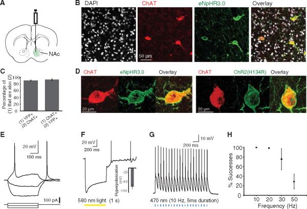
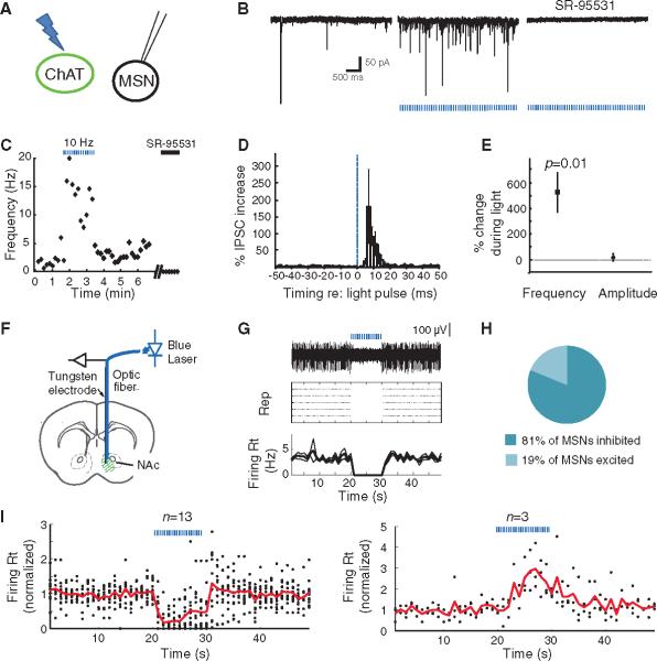
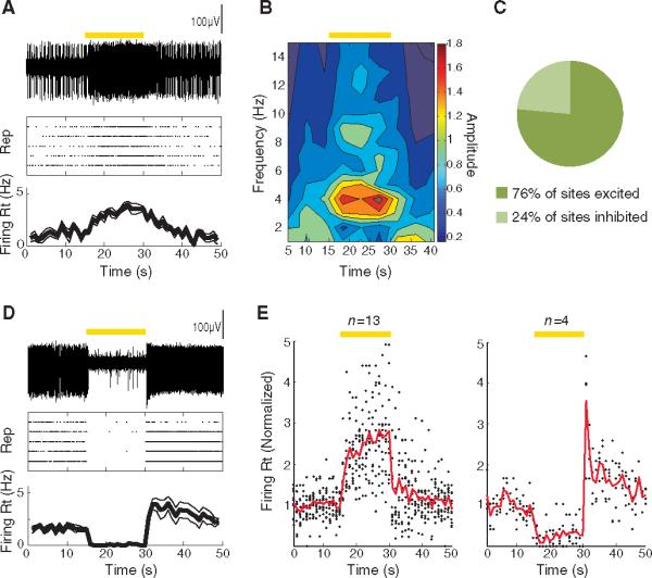
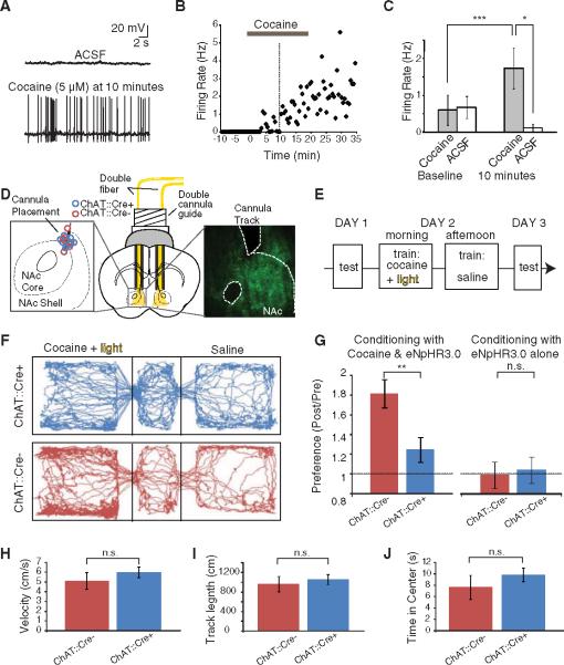
Cholinergic neurons are widespread, and pharmacological modulation of acetylcholine receptors affects numerous brain processes, but such modulation entails side effects due to limitations in specificity for receptor type and target cell. As a result, causal roles of cholinergic neurons in circuits have been unclear. We integrated optogenetics, freely moving mammalian behavior, in vivo electrophysiology, and slice physiology to probe the cholinergic interneurons of the nucleus accumbens by direct excitation or inhibition. Despite representing less than 1% of local neurons, these cholinergic cells have dominant control roles, exerting powerful modulation of circuit activity. Furthermore, these neurons could be activated by cocaine, and silencing this drug-induced activity during cocaine exposure (despite the fact that the manipulation of the cholinergic interneurons was not aversive by itself) blocked cocaine conditioning in freely moving mammals.


Figures

Fig. 1
Fig. 1
Specificity, membrane targeting, and functionality of ChR2 and eNpHR3.0 in ChAT inter-neurons of the NAc. (A) Cre-dependentAAV[expressing either eNpHR3.0-eYFP or ChR2(H134R)-eYFP] was injected into the medial portion of the NAc.(B) Confocal image of an injected slice demonstrates colocalization of eYFP expression with the ChAT antibody, costained with 4′,6′-diamidino-2-phenylindole (DAPI). (C) 91.3 ± 1.3% of neurons that expressed YFP also stained for the ChAT antibody (n = 418); 93.5 ± 2.8% of neurons that stained for the ChAT antibody also expressed YFP (n = 413). Error bars indicate SEM. (D)High-magnification view reveals membrane localization of eNphR3.0-eYFP (left) and ChR2-eYFP (right), costained with ChAT antibody. (E) Membrane potential changes induced by current injection in a ChR2-eYFP-expressing ChAT neuron. VM = −48 mV. Current steps: −60, −20, +20 pA. (F) Membrane potential changes induced by 1 s of 580-nm light in an eNpHR3.0-eYFP-expressing ChAT neuron (peak hyperpolarization: −103 mV). VM = −49 mV. (Inset) Population-averaged peak hyperpolarization (mean ± SEM: −83.8 ± 11.9 mV; n =4). (G) Consecutive action potentials in a ChR2-eYFP-expressing ChAT neuron evoked by a 470-nm pulse train (5 ms pulse width;10Hz).(H) Average success probability for generating action potentials in ChR2-eYFP-expressing ChAT neurons at different stimulation frequencies (n = 4; mean ± SEM; 470-nm pulse train, 5-ms pulse width).
Fig. 2
Fig. 2
Optogenetic photoactivation of ChAT interneurons increases frequency of inhibitory currents and suppresses MSN spiking. (A)ChAT neurons transduced with ChR2-eYFP were activated with blue light (470 nm) in brain slices, and nearby MSNs (eYFP cells) were whole-cell patch-clamped. (B) (Left) Spontaneous synaptic currents were observed in an MSN in a slice expressing ChR2-eYFP in ChAT neurons. (Middle) Synaptic currents increased in frequency in response to 470-nm light pulses (5-ms pulse width; 10 Hz). (Right) These currents were blocked by GABAA receptor antagonist SR-95531 (5 mM) and are thus considered IPSCs. 2,3-dihydroxy-6-nitro-7-sulfamoylbenzo[f]quinoxaline-2,3-dione (NBQX) (5 mM) and (RS)-3-(2-Carboxypiperazin-4-yl)-propyl-1-phosphonic acid (RS-CPP) (5 μM) were present in all experiments. (C)Time course of IPSC frequencies for this MSN, showing the effect of light pulses (blue dashed bars) and SR-95531 (black bar). (D)Average percentage increase in IPSC frequency during the light-on periods (normalized to that of light-off periods) as a function of time relative to light pulses (n = 6). The blue dashed line indicates the onset of light pulses; error bars denote SEM. (E) Light pulses increased the frequency of IPSCs by 525.8 ± 154.3% (n =6, P = 0.01, paired two-tailed t test), whereas the average amplitudes of spontaneous IPSCs were changed by 21.3 ± 28.9% (P >0.05). (F) An optrode (optical fiber attached to a tungsten electrode) was stereotaxically positioned in vivo into a NAc that expressed ChR2-eYFP in ChAT cells. (G) (Top) Voltage trace of an isolated unit that is inhibited by blue light stimulation. (Middle) Raster plot displaying the response of the same unit to five repetitions of the light stimulation, with each action potential represented by a dot. (Bottom) Average and SEM of the firing rate over time for the same unit. (H) Fraction of sites that were inhibited versus excited by light stimulation. (I) Population summary of the time course of response to light stimulation for sites that were inhibited (left; n = 13 of 16) or excited (right; n =3 of 16)by light. Solid lines represent average firing rate across sites as a function of time; each dot represents the average firing rate of an individual site. All firing rates are normalized to the mean rate before light stimulation. (F to I) Duration of photostimulation, 10 s; pulse duration, 5 ms; wavelength, 470 nm; frequency, 10 Hz. Epochs of light stimulation are represented by blue dashed lines.
Fig. 3
Fig. 3
Optogenetic photoinhibition of ChAT interneurons enhances MSN spiking in vivo. (A) (Top) Voltage trace of an isolated unit (recorded from the NAc in vivo) that was excited by optogenetic photoinhibition of the ChAT interneurons with eNpHR3.0. (Middle) Raster plot displaying the response of the same unit to five repetitions of the light stimulation, with each action potential represented by a dot. (Bottom) Average and SEM of the firing rate over time for the same unit. (B) Wavelet analysis reveals power of spiking as a function of frequency and time (average across five repetitions) for the same unit as in (A). (C) Fraction of sites that were inhibited versus excited by light stimulation. (D) Sameas (A), for a unit that was inhibited by light stimulation. (E) Population summary of the time course of response to light stimulation for sites that were inhibited (left; n = 13 of 17) or excited (right; n = 4 of 17) by light. Solid lines represent the average firing rate across sites as a function of time; each dot represents the average firing rate of an individual site. All firing rates are normalized to the mean value before light stimulation. (A to E) Duration of photostimulation, 15 s (constant illumination); wavelength, 560 nm. Epochs of light stimulation are represented by yellow bars.
Fig. 4
Fig. 4
ChAT interneurons can be activated by cocaine in slice and required for cocaine conditioning in vivo. (A) The frequency of spontaneous action potentials in a ChAT neuron increased 10 min after bath application of cocaine (5 μM). ACSF, artificial cerebrospinal fluid. (B) Firing rate over time for this ChAT neuron. Horizontal gray bar, application of cocaine; vertical dotted line, 10 min after cocaine application, the time point illustrated in detail in (A) and (C). (C) Population data illustrating the cocaine-induced increase in firing in ChAT neurons, comparing the baseline firing rate (averaged over the 2.5 min before cocaine application) with the rate after cocaine infusion (averaged between 10 and 12.5 min after onset of cocaine application; gray bars, cells receiving cocaine; white bars, control cells receiving only ACSF; P < 0.005, paired two-tailed t test for cocaine-treated group before versus after cocaine; P < 0.05 unpaired two-tailed t test comparing cocaine versus control cells after cocaine or vehicle). (D) Schematic illustration of a bilateral cannula system with double fibers inserted to illuminate the medial portion of the NAc. (Left inset) Endpoint of cannula track for all mice used in (H). (Right inset) eYFP expression in NAc of a ChAT∷Cre+ mouse injected with Cre-dependent eNpHR3.0-eYFP. (E) Conditioning paradigm for cocaine CPP (H). Mice were conditioned with ip cocaine (20 mg/kg), along with ChAT cell inhibition with eNpHR3.0 (wavelength: 590 nm). (F) Tracking data from representative ChAT∷Cre+ and ChAT∷Cre mice on the testing day after cocaine conditioning (day 3). On the previous day (day 2), the mice had received cocaine and light in one left chamber, whereas in the other they received saline. The ChAT∷Cre mouse (but not the ChAT∷Cre+ mouse) exhibited a preference for the conditioned chamber. (G) (Left) Fold change in time in conditioned chamber during day 3 versus day 1 of cocaine CPP (conditioning with cocaine and light). Comparison of ChAT∷Cre+ and ChAT∷Cre littermates; in both cases injected with Cre-dependent eNpHR3.0 (n = 10 ChAT∷Cre+, n = 12 ChAT∷Cre; P < 0.01 for two-tailed t test; three cohorts). (Right) Fold change in time in conditioned chamber during day 3 versus day 1 for conditioning with light alone (no cocaine; n = 9 ChAT∷Cre+, n = 7 ChAT∷Cre; P > 0.05 for two-tailed t test; three cohorts). Error bars indicate SEM. n.s., not significant. (H) Velocity of virus-injected (Cre-dependent eNpHR3.0) and photostimulated ChAT∷Cre+ and ChAT∷Cre mice in the open field (n = 10 ChAT∷Cre+, n = 10 ChAT∷Cre; P > 0.05 for two-tailed t test; three cohorts). (I) Same as (H) for track length in open field (n = 10 ChAT∷Cre+, n = 10 ChAT∷Cre; P > 0.05 for two-tailed t test; three cohorts). (J) Same as (H) for time in center of open field (n = 10 ChAT∷Cre+, n = 10 ChAT∷Cre; P > 0.05 for two-tailed t test; three cohorts). (A to J) *P < 0.05; **P < 0.01; ***P < 0.005.

References

    1. Changeux JP. C. R. Biol. 2009;332:421. - PubMed
    1. Kilgard MP, Merzenich MM. Science. 1998;279:1714. - PubMed
    1. Bakin JS, Weinberger NM. Proc. Natl. Acad. Sci. U.S.A. 1996;93:11219. - PMC - PubMed
    1. Maskos U. Br. J. Pharmacol. 2008;153(suppl. 1):S438. - PMC - PubMed
    1. Picciotto MR, et al. Nature. 1998;391:173. - PubMed

Publication types

MeSH terms