Review
. 2011 Feb;22(2):216-24.
doi: 10.1681/ASN.2010020186. Epub 2010 Dec 16.

The regulation of parathyroid hormone secretion and synthesis

Affiliations
Review

The regulation of parathyroid hormone secretion and synthesis

Rajiv Kumar et al. J Am Soc Nephrol. 2011 Feb.

Abstract

Secondary hyperparathyroidism classically appears during the course of chronic renal failure and sometimes after renal transplantation. Understanding the mechanisms by which parathyroid hormone (PTH) synthesis and secretion are normally regulated is important in devising methods to regulate overactivity and hyperplasia of the parathyroid gland after the onset of renal insufficiency. Rapid regulation of PTH secretion in response to variations in serum calcium is mediated by G-protein coupled, calcium-sensing receptors on parathyroid cells, whereas alterations in the stability of mRNA-encoding PTH by mRNA-binding proteins occur in response to prolonged changes in serum calcium. Independent of changes in intestinal calcium absorption and serum calcium, 1α,25-dihydroxyvitamin D also represses the transcription of PTH by associating with the vitamin D receptor, which heterodimerizes with retinoic acid X receptors to bind vitamin D-response elements within the PTH gene. 1α,25-Dihydroxyvitamin D additionally regulates the expression of calcium-sensing receptors to indirectly alter PTH secretion. In 2°HPT seen in renal failure, reduced concentrations of calcium-sensing and vitamin D receptors, and altered mRNA-binding protein activities within the parathyroid cell, increase PTH secretion in addition to the more widely recognized changes in serum calcium, phosphorus, and 1α,25-dihydroxyvitamin D. The treatment of secondary hyperparathyroidism by correction of serum calcium and phosphorus concentrations and the administration of vitamin D analogs and calcimimetic agents may be augmented in the future by agents that alter the stability of mRNA-encoding PTH.


Figures

Figure 1
Figure 1
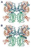
A hypothetical dimeric model of residues D23 (blue) to I528 (red) of the human calcium sensing receptor extracellular domain (CaSR ECD). (A) Both monomers containing just the Venus flytrap region of the CaSR ECD are shown in a closed and presumably active conformation as was reported for the extracellular domain of the glutamate receptor with glutamate bound. The two yellow spheres (yellow arrows) indicate putative Ca2+-binding sites, found at the nexus of where both lobes of a monomer meet. Most residues forming this cation-binding site are not conserved in glutamate receptor. The additional cyan spheres within the topmost lobes of the dimer designate possible Mg2+-binding sites (green spheres indicated by green arrows) brought over from glutamate receptor coordinates. These Mg2+ sites are completely conserved in CaSR. The dimer interface of the portion of CaSR shown is completely formed from interactions between these two upper lobes. There are no intermolecular disulfide bridges linking the dimer together within this portion of the ECD of CaSR, although two intramolecular disulfides exist. (B) A model of the apo-CaSR dimer is portrayed. Again, the color ramps from blue to red from D23 to I528. The Mg2+ sites are present, although there is no experimental basis for this premise. Of note is the significant opening and expansion of the cavities between the upper and lower lobes of each monomer, the areas indicated by the two yellow ovals. (C) The upper lobes of the CaSR atomic coordinates shown above in (A) (with Ca2+ bound, now made gray in color) are superimposed on the apo-form model for the CaSR dimer drawn in rainbow as in (B). The red arrows point to a large displacement in the orientation and position of the carboxyterminal end of the structure near where the CaSR cysteine-rich domains (not shown) might be found. Significant conformational changes within parts of the CaSR ECD connecting with the transmembrane domains probably occur on Ca binding.
Figure 2
Figure 2
Models of bound phenylalanine and neomycin molecules within the cavities of the CaSR dimer. (A) Above the predicted Ca2+-binding sites shown by yellow spheres are phenylalanine molecules shown in a conformation that stacks its side-chain ring against a tryptophan residue that is unique to CaSR, whereas remaining atoms occupy the same locations as found for the glutamate molecules bound to glutamate receptor. (B) Two neomycin molecules may also be docked within a third buried location as shown in the bottom-most image.
Figure 3
Figure 3
Pathways by which the CaSR homodimer signals in cells after binding of Ca2+ to the extracellular domains (red line) of the CaSR molecules in the homodimeric pair. Through the association of the CaSR with the i-type heterotrimeric G protein, Giα, adenylate cyclase (AC) activity is inhibited and cyclic AMP (cAMP) concentrations decrease. Association of the CaSR with the Gqα subunit of q-type heterotrimeric G protein results in the activation of PLC that increases inositol (1,4,5)P3 and diacylglycerol (DAG) with attendant downstream effects such as an increase in intracellular calcium that is mobilized from intracellular stores, and the activation of PKC. MAPK and PLA2 are activated by Gqα-dependent pathways with increases in MEK and ERK and an increase in arachidonic acid formation.
Figure 4
Figure 4
(A) Cellular processing of mRNA. Nascent mRNA comprised of exons (E1 through E4) and intervening sequences (IVS) is processed in the nucleus by 5′-methyl capping, splicing, cleavage, and polyadenylation. In the cytoplasm, AU-rich element–binding proteins (ARE-BPs, blue box and red oval) bind to AREs within the 3′-region of RNA and stabilize or destabilize mRNA. Stabilized mRNA undergoes translation in ribosomes, whereas destabilized mRNA undergoes deadenylation, decapping, and degradation in exosomes or P-bodies. (Adapted from reference with permission from the American Society for Clinical Investigation.) (B) Processing of mRNA-encoding PTH. Murine mRNA-encoding PTH is bound by ARE-BPs, which either stabilize or destabilize the mRNA. The ratio of activities of stabilizing/destabilizing ARE-binding proteins bound to mRNA-encoding PTH determines the half-life of the mRNA. KSRP is a mRNA-destabilizing ARE-BP for mRNA-encoding PTH that is active in its dephosphorylated state. The peptidyl-prolyl isomerase Pin1 is responsible for the dephosphorylation of KSRP. In CKD, Pin1 activity is reduced, and as a result less dephosphorylated (active) KSRP is available. Consequently, a stabilizing ARE-BP, AUF1, is active and mRNA-encoding PTH is degraded to a lesser extent, resulting in higher intracellular mRNA levels, more PTH synthesis, and secondary hyperparathyroidism. Abbreviation: P, phosphate. (Adapted from reference with permission from the American Society for Clinical Investigation.)
Figure 5
Figure 5
Alterations within the parathyroid gland that favor the development of 2°HPT in the context of CRF and ESRD.

References

    1. Habener JF, Rosenblatt M, Potts JT., Jr Parathyroid hormone: Biochemical aspects of biosynthesis, secretion, action, and metabolism. Physiol Rev. 1984;64:985–1053. - PubMed
    1. Potts JT. Parathyroid hormone: Past and present. J Endocrinol. 2005;187:311–325. - PubMed
    1. Kumar R. Metabolism of 1,25-dihydroxyvitamin D3. Physiol Rev. 1984;64:478–504. - PubMed
    1. DeLuca HF, Schnoes HK. Vitamin D: Recent advances. Annu Rev Biochem. 1983;52:411–439. - PubMed
    1. Gesek FA, Friedman PA. On the mechanism of parathyroid hormone stimulation of calcium uptake by mouse distal convoluted tubule cells. J Clin Invest. 1992;90:749–758. - PMC - PubMed

Publication types

MeSH terms