. 2011 Apr;18(4):690-9.
doi: 10.1038/cdd.2010.163. Epub 2010 Dec 17.

Anti-apoptotic effect of hyperglycemia can allow survival of potentially autoreactive T cells

Affiliations

Anti-apoptotic effect of hyperglycemia can allow survival of potentially autoreactive T cells

P Ramakrishnan et al. Cell Death Differ. 2011 Apr.

Erratum in

  • Cell Death Differ. 2012 Nov;19(11):1892

Abstract

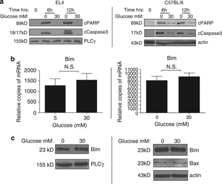
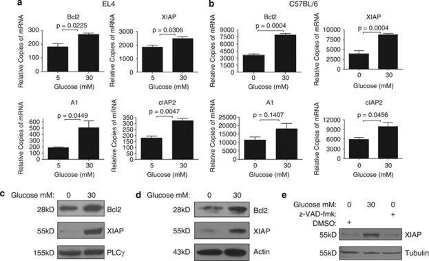
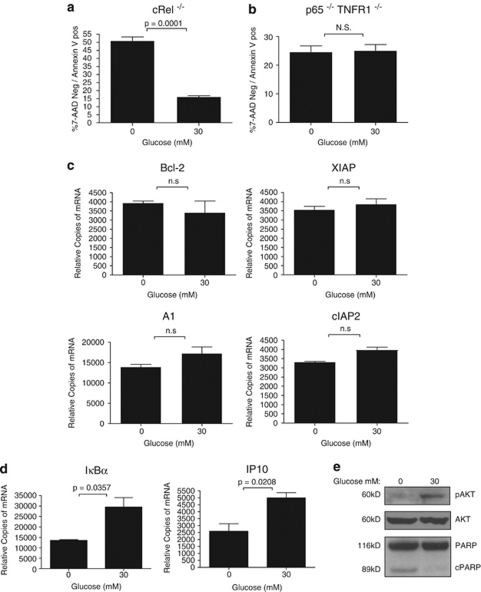
Thymocyte development is a tightly controlled multi-step process involving selective elimination of self-reactive and non-functional T cells by apoptosis. This developmental process depends on signaling by Notch, IL-7 and active glucose metabolism. In this study, we explored the requirement of glucose for thymocyte survival and found that in addition to metabolic regulation, glucose leads to the expression of anti-apoptotic genes. Under hyperglycemic conditions, both mouse and human thymocytes demonstrate enhanced survival. We show that glucose-induced anti-apoptotic genes are dependent on NF-κB p65 because high glucose is unable to attenuate normal ongoing apoptosis of thymocytes isolated from p65 knockout mice. Furthermore, we demonstrate that in vivo hyperglycemia decreases apoptosis of thymocytes allowing for survival of potentially self-reactive thymocytes. These results imply that hyperglycemic conditions could contribute to the development of autoimmunity through dysregulated thymic selection.


Figures

Figure 1
Figure 1
(a) EL-4 cells are unique in glucose dependence for survival. Equivalent numbers of starting cells (1 × 10⁶ per well for the suspension cells in 12-well plate and 1.5 × 10⁵ per well in six-well plate for adherent cells) were seeded in media lacking glucose after having been growing in standard glucose containing media (RPMI 11.1 mM glucose and DMEM 25 mM glucose). After 24 h, cell numbers were determined by Trypan blue exclusion. Data are representative of three independent experiments. (be) EL-4 cells are dependent on glucose for survival and proliferation. EL-4 cell cultures seeded with equivalent numbers of starting cells (1 × 10⁶) in 12-well plate in RPMI with indicated final concentrations of glucose or mannitol. After 20 h, cell number determined by Trypan blue exclusion (b) and 24 h later by visual inspection on a Nikon Diaphot 300 phase contrast microscope (NIKON, Melville, NY, USA) at × 400 final magnification and photographed using a SPOT digital camera and software (Sterling Heights, MI USA). (d) Evaluation of EL-4 cells as described above at 12 h showed apoptotic morphology. (e) EL-4 cells were cultured at the indicated glucose concentrations for 12 h and then evaluated by FACS for evidence of early apoptosis (7-AAD negative/Annexin V positive). Data are representative of four independent experiments
Figure 2
Figure 2
Enhanced thymocyte glucose-mediated survival is restricted to the CD4+CD8+ population. (a and b) Single-cell suspensions were prepared from male C57BL/6 thymuses and cultured for 24 h in vitro with the indicated concentration of glucose. The cell suspensions were collected and stained for CD4, CD8, 7-AAD and Annexin V for FACS evaluation. Indicated sub-populations were evaluated for cells in the early stages of apoptosis (7-AAD negative/Annexin V positive). (c) Composite thymic subpopulation evaluations from male C57BL/6 (n=4). (d) Single-cell suspension was prepared from a human fetal thymus (gestational age=16 weeks) isolated immediately following the surgical termination. The cell suspension was cultured for 24 h in vitro with the indicated concentration of glucose, subsequently stained for CD4, CD8, 7-AAD and Annexin V for FACS evaluation for early stage apoptosis (7-AAD negative/Annexin V positive). Statistical difference determined by two-tailed unpaired Student's t-test (N.S., nonsignificant)
Figure 3
Figure 3
Glucose inhibits thymocyte caspase activation, but not through altered tonic Bim levels. (a) EL-4 cells (left) or single-cell suspension from male C57BL/6 thymocytes were cultured for the indicated times and glucose concentrations before total cellular protein isolation. Western blot membranes were probed for cleaved PARP (cPARP), cleaved caspase 3 (cCaspase3). Phospholipase C γ (PLCγ) and actin were used as the loading controls. (b) Total RNA was isolated from EL-4 cells (left) or single-cell suspension from male C57BL/6 thymocytes that had been cultured for the 6 h at indicated glucose concentrations. As the EL4 cells were unable to survive at 0 mM glucose, the cells were cultured overnight at 5 mM glucose and then treated with 30 mM glucose. Quantitative RT-PCR was performed in triplicate and relative abundance of Bim transcripts calculated. Statistical difference determined by two-tailed unpaired Student's t-test (N.S., nonsignificant). (c) Total cellular proteins were isolated as above. Western blot membranes from EL-4 cells were probed for Bim and PLCγ (left). Western blot membranes from C57BL/6 thymocytes were probed from Bim and Bax
Figure 4
Figure 4
High glucose causes upregulation of anti-apoptotic genes. Total RNA was isolated from EL-4 cells (a) or single-cell suspension from male C57BL/6 thymus (b) grown and treated as in Figure 3b. Quantitative RT-PCR was performed in triplicate and relative abundance of Bcl2, XIAP, A1 and cIAP transcripts calculated. Statistical difference determined by two-tailed unpaired Student's t-test (NS, nonsignificant). EL-4 cells (c) or single-cell suspension from male C57BL/6 (d) were cultured for 10 and 24 h, respectively at the indicated glucose concentrations before total cellular protein isolation. Western blot membranes were probed for Bcl2 and XIAP. Actin and Phospholipase Cγ (PLCγ) were used as loading controls. (e) EL-4 cells were cultured for 10 h under the indicated conditions (z-VAD-fmk at a final concentration of 50 μM) before total cellular protein isolation. Western blot membranes were probed for XIAP and tubulin was used as a loading control
Figure 5
Figure 5
Anti-apoptotic glucose effect on thymocytes is dependent on p65 NF-κB. Single-cell suspensions were prepared from (a) male c-Rel−/− C57BL/6 (n=4) or (b) male p65−/− TNFR1−/− (n=2) thymuses and cultured for 24 h in vitro with the indicated concentration of glucose. The cell suspensions were collected and stained for CD4, CD8, 7-AAD and Annexin V for FACS evaluation. CD4+CD8+ population was evaluated for early stages of apoptosis (7-AAD negative/Annexin V positive). Statistical difference determined by two-tailed unpaired Student's t-test (N.S., nonsignificant). (c) Total RNA was isolated from male p65−/− TNFR1−/− thymic single-cell suspension cultured for 6 h at the indicated glucose concentrations. Quantitative RT-PCR was performed in triplicate and relative abundance of Bcl2, XIAP, A1 and cIAP transcripts calculated. Statistical difference determined by two-tailed unpaired Student's t-test (N.S., nonsignificant). (d and e) High glucose induces general activation of the NF-κB pathway and phosphorylation of AKT (pAKT). (d) Total RNA was isolated from single-cell suspension cultured in vitro at the indicated glucose concentrations from male C57BL/6 thymus and the relative abundance of IκBα and IP10 transcripts determined by qPCR. (e) Total protein was isolated from the thymocyte cultures above and western blot was probed for pAKT, AKT and PARP
Figure 6
Figure 6
Hyperglycemia results in reduced deletion of antigen-specific transgenic TCR thymocytes when challenged with antigen. C57BL/6 and C57BL/6 – OT2 were treated with STZ (n=4) or saline (n=4). After 3 weeks, the mice were given 10 mg/kg ovalbumin i.p. The mice were euthanized 24 h later. Blood from cardiac puncture was evaluated for glucose concentration (a). Thymic single-cell suspensions were created from the above mice, surface stained for CD4 and CD8, and sub-populations (bd) evaluated by FACS. Statistical difference determined by two-tailed unpaired Student's t-test (N.S., nonsignificant)

Similar articles

Cited by

References

    1. Patterson CC, Dahlquist GG, Gyurus E, Green A, Soltesz G. Incidence trends for childhood type 1 diabetes in Europe during 1989–2003 and predicted new cases 2005–20: a multicentre prospective registration study. Lancet. 2009;373:2027–2033. - PubMed
    1. Ciofani M, Zuniga-Pflucker JC. Notch promotes survival of pre-T cells at the beta-selection checkpoint by regulating cellular metabolism. Nat Immunol. 2005;6:881–888. - PubMed
    1. Hayday AC, Pennington DJ. Key factors in the organized chaos of early T cell development. Nat Immunol. 2007;8:137–144. - PubMed
    1. Zhang N, Hartig H, Dzhagalov I, Draper D, He YW. The role of apoptosis in the development and function of T lymphocytes. Cell Res. 2005;15:749–769. - PubMed
    1. Greiner EF, Guppy M, Brand K. Glucose is essential for proliferation and the glycolytic enzyme induction that provokes a transition to glycolytic energy production. J Biol Chem. 1994;269:31484–31490. - PubMed

Publication types

MeSH terms

Substances