

What should be considered in treatment of melasma
- PMID: 21165205
- PMCID: PMC2991712
- DOI: 10.5021/ad.2010.22.4.373
What should be considered in treatment of melasma
Abstract
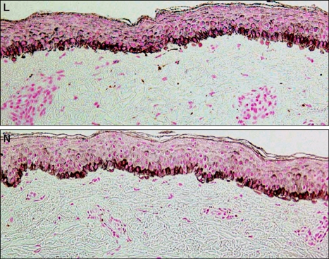
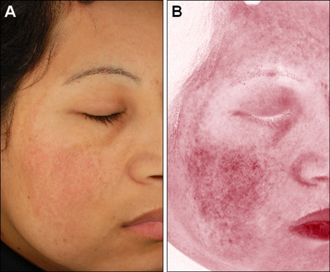
Melasma is a common acquired hyperpigmentary skin disorder characterized by light to dark brown macules and patches occurring in the sun-exposed areas of the face. Melasma lesional skin is characterized by epidermal hyperpigmentation through increased melanogenesis in epidermal melanocytes. Some patients have dermal melanin but its amount is not significant and its distribution is very heterogeneous in the whole melasma lesional skin. Melasma is not homogeneous disease and there are personal characteristics of patients with melasma. The pathogenesis of melasma is not fully understood, but several hypotheses have been suggested. Increased vascularity in melasma lesions has suggested the role of increased number of enlarged vessels in the development of melasma. Endogeneous and exogeneous stimuli such as sex hormones and ultraviolet irradiation respectively may stimulate the microenvironment leading to the release of various mediators that cause activation of melanocytes and/or these stimuli may directly activate the melanocytes. Melasma patients may have specialized melanocytes with an intrinsic sensitivity to these stimuli.
Keywords: Melanin; Melanocytes; Melasma; Vascularity.
Conflict of interest statement
The authors have no conflicts of interest that are directly relevant to the content of this review.
Figures
References
-
- Ortonne JP, Arellano I, Berneburg M, Cestari T, Chan H, Grimes P, et al. A global survey of the role of ultraviolet radiation and hormonal influences in the development of melasma. J Eur Acad Dermatol Venereol. 2009;23:1254–1262. - PubMed
-
- Kang HY, Valerio L, Bahadoran P, Ortonne JP. The role of topical retinoids in the treatment of pigmentary disorders: an evidence-based review. Am J Clin Dermatol. 2009;10:251–260. - PubMed
-
- Ortonne JP, Passeron T. Melanin pigmentary disorders: treatment update. Dermatol Clin. 2005;23:209–226. - PubMed
-
- Negishi K, Kushikata N, Tezuka Y, Takeuchi K, Miyamoto E, Wakamatsu S. Study of the incidence and nature of "very subtle epidermal melasma" in relation to intense pulsed light treatment. Dermatol Surg. 2004;30:881–886. - PubMed
-
- Lee HS, Won CH, Lee DH, An JS, Chang HW, Lee JH, et al. Treatment of melasma in Asian skin using a fractional 1,550-nm laser: an open clinical study. Dermatol Surg. 2009;35:1499–1504. - PubMed
LinkOut - more resources
Full Text Sources
Other Literature Sources
