. 2010 Oct;14(5):317-24.
doi: 10.4196/kjpp.2010.14.5.317. Epub 2010 Oct 31.

Regional Distribution of Interstitial Cells of Cajal (ICC) in Human Stomach

Affiliations

Regional Distribution of Interstitial Cells of Cajal (ICC) in Human Stomach

Hyo-Yung Yun et al. Korean J Physiol Pharmacol. 2010 Oct.

Abstract

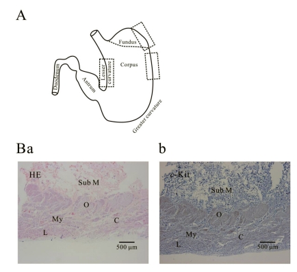
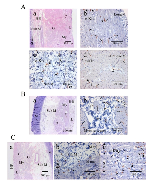
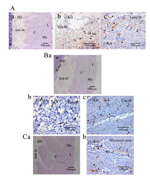
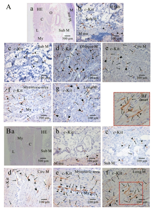
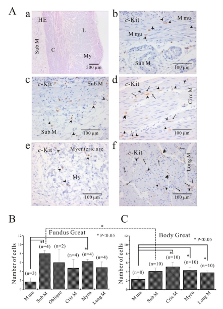
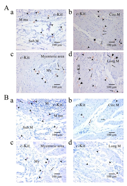
We elucidated the distribution of interstitial cells of Cajal (ICC) in human stomach, using cryosection and c-Kit immunohistochemistry to identify c-Kit positive ICC. Before c-Kit staining, we routinely used hematoxylin and eosin (HE) staining to identify every structure of human stomach, from mucosa to longitudinal muscle. HE staining revealed that the fundus greater curvature (GC) had prominent oblique muscle layer, and c-Kit immunostaining c-Kit positive ICC cells were found to have typical morphology of dense fusiform cell body with multiple processes protruding from the central cell body. In particular, we could observe dense processes and ramifications of ICC in myenteric area and longitudinal muscle layer of corpus GC. Interestingly, c-Kit positive ICC-like cells which had morphology very similar to ICC were found in gastric mucosa. We could not find any significant difference in the distribution of ICC between fundus and corpus, except for submucosa where the density of ICC was much higher in gastric fundus than corpus. Furthermore, there was no significant difference in the density of ICC between each area of fundus and corpus, except for muscularis mucosa. Finally, we also found similar distribution of ICC in normal and cancerous tissue obtained from a patient who underwent pancreotomy and gastrectomy. In conclusion, ICC was found ubiquitously in human stomach and the density of ICC was significantly lower in the muscularis mucosa of both fundus/corpus and higher in the submucosa of gastric fundus than corpus.

Keywords: Corpus; Fundus; Greater and Lesser curvature; Human stomach; Interstitial cells of Cajal (ICC); c-Kit.


Figures

Fig. 1
Fig. 1
Histology of human gastric wall. As shown in panel A, we used fundus greater curvature, corpus greater curvature and corpus lesser curvature of human stomach. In HE staining of fundus greater curvature (Ba), we identified oblique, circular and longitudinal muscles. And then in c-Kit immunostaining (Bb), we observed c-Kit positive immnuhistochemical reactivity of ICC, which appeared as dark brown spots. Sub M, submucosa; O, oblique muscle; C, circular muscle; My, myenteric region; L, longitudinal muscle.
Fig. 2
Fig. 2
Distribution of ICC in human gastric fundus of greater curvature. In HE staining (Aa, Ba, Ca), the whole layers of human gastric fundus from greater curvature can be seen. Oblique muscle layer is prominent in human gastric fundus. In c-Kit immunostaining, we identified c-Kit positive ICC in every muscle layers: longitudinal muscle (Ab), submucosa (Ac), oblique muscle (Ad), myenteric area (Bb), muscularis mucosa (Cb) and circular muscle (Cc). Since we used cryosection for immunohistochemical study, ICC could not be seen as a whole structure. Instead, ICC is seen in long spindle-like (fusiform) shaped cell body and multiple processes from the central cell body, which is especially prominent in (Ab, Ad, Bb). ICC is identified in submucosa, muscularis mucosa and mucosa, too (Ac) and (Cb). Arrow: processes or ramification, arrow head: central cell body, double arrow head: ICC in septa (ICC-SEP).
Fig. 3
Fig. 3
Distribution of ICC in human gastric corpus of greater curvature. In HE staining (Aa, Ba, Ca), the whole layers of human gastric corpus from greater curvature can be seen. In c-Kit immunostaining, c-Kit positive ICC is also observed in every layer; muscularis mucosa (Ab), longitudinal muscle (Ac), submucosa (Bb), circular muscle (Bc), and myenteric area (Cb). In human gastric corpus, oblique muscle is not as prominent as in fundus. ICC is seen in long spindle-like (fusiform) shaped cell body and multiple processes from the central cell body, and is also identified in mucosa (Ab). Arrow: processes or ramification, arrow head: central cell body.
Fig. 4
Fig. 4
Comparison of histology between fundus and corpus in greater curvature of human stomach. In HE staining, the whole layer of fundus (Aa) and corpus (Ba) are shown. It is striking to find that fundus has prominent structure of oblique muscle, unlike corpus. c-Kit positive ICC is observed in every layer of human gastric fundus (Ab~Ag) and corpus (Bb~Bf) of greater curvature. Interestingly, almost all kinds of ICC are found in oblique muscle layer, however, ICCs of circular and longitudinal muscle are ICC-SEP. In panel (Bf and Bf inset), it is evident that ICC of longitudinal muscle has lots of processes and ramifications. Arrow: processes or ramification, arrow head: central cell body, double arrow head: ICC in septa (ICC-SEP).
Fig. 5
Fig. 5
Comparison of ICC distribution between corpus and fundus and regional distribution of ICC in part of human stomach. In HE staining (Aa), the whole layers of human gastric corpus from lesser curvature are demonstrated. In corpus lesser curvature, c-Kit positive ICC is also found in all the layers (A). ICC numbers are compared between fundus greater curvature (B) and corpus greater curvature (C). There is no significant difference between two regions except for ICC from submucosa. The average number of ICC is 8±0.7 count/unit area in submucosa of fundus greater curvature, which is significantly higher than 4.1±0.7 count/unit area of body greater curvature (p<0.05). Meanwhile, the density of ICC in muscularis mucosa is significantly less than that of other layers of fundus (submucosa and myenteric area) and corpus (circular muscle, myenteric area and longitudinal muscle) (p<0.05; (5B) and (5C)). However, the density of ICC in muscularis mucosa is less than that of other layer with no significant difference (p>0.05; (5B) and (5C)). Arrow: processes or ramification, arrow head: central cell body, double arrow head: ICC in septa (ICC-SEP).
Fig. 6
Fig. 6
Comparison of ICC distribution between normal and cancerous tissue of human stomach. Distribution of ICC is compared between normal (A) and cancerous tissue (B) obtained from a patient who underwent pancreotomy and gastrectomy. c-Kit positive ICC is observed in every layer; submucosa (Aa, Ba), circular muscle (Ab, Bb), myenteric area (Ac, Bc), and longitudinal muscle (Ad, Bd). Note that c-Kit positive immunoreactivity is found also in gastric mucosa of cancer area (Ba) as well as normal area (Aa). Arrow: processes or ramification, arrow head: central cell body, double arrow head: ICC in septa (ICC-SEP).

References

    1. Cajal SR. Histologie du système nerveux de l'homme et des vertébrés. 2nd ed. Paris: Maloine; 1911. pp. 891–942.
    1. Faussone-Pellegrini MS, Cortesini C. Ultrastructural features and localization of the interstitial cells of Cajal in the smooth muscle coat of human esophagus. J Submicrosc Cytol. 1985;17:187–197. - PubMed
    1. Hagger R, Gharaie S, Finlayson C, Kumar D. Regional and transmural density of interstitial cells of Cajal in human colon and rectum. Am J Physiol. 1998;275:G1309–G1316. - PubMed
    1. Radenkovic G, Savic V, Mitic D, Grahovac S, Bjelakovic M, Krstic M. Development of c-Kit immunopositive interstitial cells of Cajal in the human stomach. J Cell Mol Med. 2010;14:1125–1134. - PMC - PubMed
    1. Romert P, Mikkelsen HB. c-Kit immunoreactive cells of Cajal in the human small and large intestine. Histochem Cell Biol. 1998;109:195–202. - PubMed