. 2010 Dec;10(4):176-89.
doi: 10.1007/s10558-010-9109-9.

On the mechanics underlying the reservoir-excess separation in systemic arteries and their implications for pulse wave analysis

Affiliations

On the mechanics underlying the reservoir-excess separation in systemic arteries and their implications for pulse wave analysis

Jordi Alastruey. Cardiovasc Eng. 2010 Dec.

Abstract

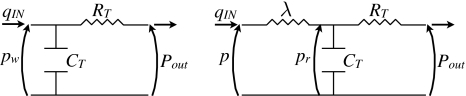
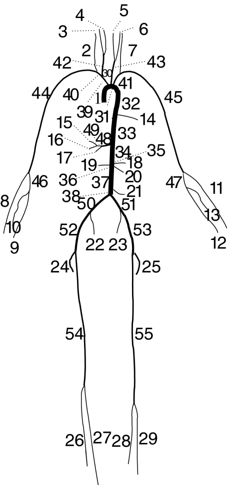
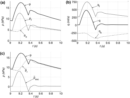
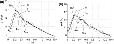
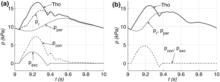
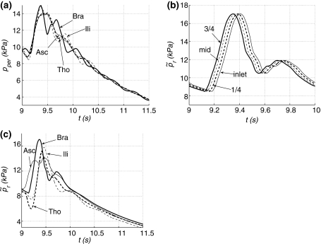
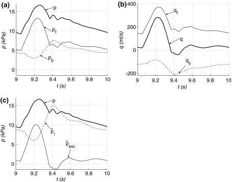
Several works have separated the pressure waveform p in systemic arteries into reservoir p(r) and excess p(exc) components, p = p(r) + p(exc), to improve pulse wave analysis, using windkessel models to calculate the reservoir pressure. However, the mechanics underlying this separation and the physical meaning of p(r) and p(exc) have not yet been established. They are studied here using the time-domain, inviscid and linear one-dimensional (1-D) equations of blood flow in elastic vessels. Solution of these equations in a distributed model of the 55 larger human arteries shows that p(r) calculated using a two-element windkessel model is space-independent and well approximated by the compliance-weighted space-average pressure of the arterial network. When arterial junctions are well-matched for the propagation of forward-travelling waves, p(r) calculated using a three-element windkessel model is space-dependent in systole and early diastole and is made of all the reflected waves originated at the terminal (peripheral) reflection sites, whereas p(exc) is the sum of the rest of the waves, which are obtained by propagating the left ventricular flow ejection without any peripheral reflection. In addition, new definitions of the reservoir and excess pressures from simultaneous pressure and flow measurements at an arbitrary location are proposed here. They provide valuable information for pulse wave analysis and overcome the limitations of the current two- and three-element windkessel models to calculate p(r).


Figures

Fig. 1
Fig. 1
Analogous electrical circuit diagrams governed by the (left) two-element windkessel Eq. 1 and (right) three-element windkessel Eq. 4
Fig. 2
Fig. 2
Connectivity of the 55 larger systemic arteries in the human, as proposed in Stergiopulos et al. (1992). Their names and properties are shown in Tables 2 and 3
Fig. 3
Fig. 3
a Pressure p and reservoir p r and excess p exc pressures calculated using the three-element windkessel model with λ = Z Ao in the midpoint of the single-vessel aortic model, and windkessel pressure formula image with time. b formula image and compliance-weighted space-average pressure p C. c Reservoir pressure p r calculated using the three-element windkessel model with λ = Z Ao at x = 0 (inlet), l/4 (1/4), l/2 (mid) and 3l/4 (3/4). d New reservoir pressure formula image at the same locations as in (c)
Fig. 4
Fig. 4
a New excess pressure formula image with time at formula image (inlet), l/4 (1/4), l/2 (mid) and 3l/4 (3/4) in the single-vessel aortic model. b Flow with time at the inlet (q IN) and midpoint (mid), and outflows formula image and q C driven by formula image and p C, respectively
Fig. 5
Fig. 5
Forward (p f and q f) and backward (p b and q b) contributions to a pressure p and b flow q, and c new reservoir formula image and excess formula image pressures with time in the midpoint of the single-vessel aortic model
Fig. 6
Fig. 6
Pressure with time at the root of the ascending aorta (Edge 1, Asc) and the midpoint of the thoracic aorta II (Edge 33, Tho), left brachial (Edge 45, Bra) and right external iliac (Edge 52, Ili) arteries of the a normal and b well-matched 55-artery models. Pressures formula image in thick solid lines. After formula image s, formula image to show the relaxation of both models
Fig. 7
Fig. 7
Pressure (Tho) with time in the midpoint of the thoracic aorta II (Edge 33) and reservoir p r (with λ = Z Ao), peripheral p per, excess p exc and conduit p con pressures at the same location in the a normal and b well-matched 55-artery models
Fig. 8
Fig. 8
a Peripheral pressure p per with time at the root of the ascending aorta (Edge 1, Asc), and the midpoint of the thoracic aorta II (Edge 33, Tho), left brachial (Edge 45, Bra) and right external iliac (Edge 52, Ili) arteries of the well-matched 55-artery model. b New reservoir pressure formula image with time at formula image (inlet), l/4 (1/4), l/2 (mid) and 3l/4 (3/4) in the left brachial artery of the normal 55-artery model. c formula image with time at Asc, Tho, Bra and Ili of the normal 55-artery model. After formula image s, formula image in panels (a) and (c)
Fig. 9
Fig. 9
a, b Windkessel formula image and compliance-weighted space-average p C pressures, and c, d outflows formula image and q C driven by formula image and p C, respectively, with time in the normal (a, c) and well-matched (b, d) 55-artery models
Fig. 10
Fig. 10
Forward (p f and q f) and backward (p b and q b) contributions to a pressure p and b flow q, and c new reservoir formula image and excess formula image pressures with time in the midpoint of the thoracic aorta II (Edge 33) of the normal 55-artery model
Fig. 11
Fig. 11
Diastolic formula image on a logarithm scale with time in the single-vessel aorta (1art) and the normal (55art) and well-matched (55art wm) 55-artery models. After formula image s, formula image to show the relaxation of the models

References

    1. Aguado-Sierra J, Alastruey J, Wang J-J, Hadjiloizou N, Davies J, Parker K. Separation of the reservoir and wave pressure and velocity from measurements at an arbitrary location in arteries. Proc Inst Mech Eng Part H J Eng Med. 2008;222:403–16. doi: 10.1243/09544119JEIM315. - DOI - PubMed
    1. Alastruey J. Numerical modelling of pulse wave propagation in the cardiovascular system: development, validation and clinical applications. Ph.D. thesis, Imperial College London, University of London, UK 2006.
    1. Alastruey J, Parker K, Peiró J, Sherwin S. Lumped parameter outflow models for 1-D blood flow simulations: effect on pulse waves and parameter estimation. Commun Comput Phys. 2008;4:317–36.
    1. Alastruey J, Parker K, Peiró J, Sherwin S. Analysing the pattern of pulse waves in arterial networks: a time-domain study. J Eng Math. 2009;64:331–51. doi: 10.1007/s10665-009-9275-1. - DOI
    1. Caro C, Pedley T, Schroter R, Seed W. The mechanics of the circulation. Oxford: Oxford University Press; 1978.

Publication types

LinkOut - more resources