Review
. 2011 Apr;4(2):138-44.
doi: 10.1007/s12265-010-9254-y. Epub 2010 Dec 17.

Circulating very small embryonic-like stem cells in cardiovascular disease

Affiliations
Review

Circulating very small embryonic-like stem cells in cardiovascular disease

Wojciech Wojakowski et al. J Cardiovasc Transl Res. 2011 Apr.

Abstract

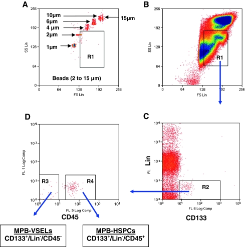
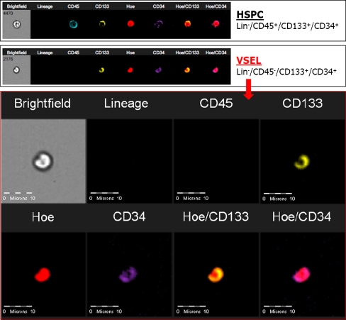
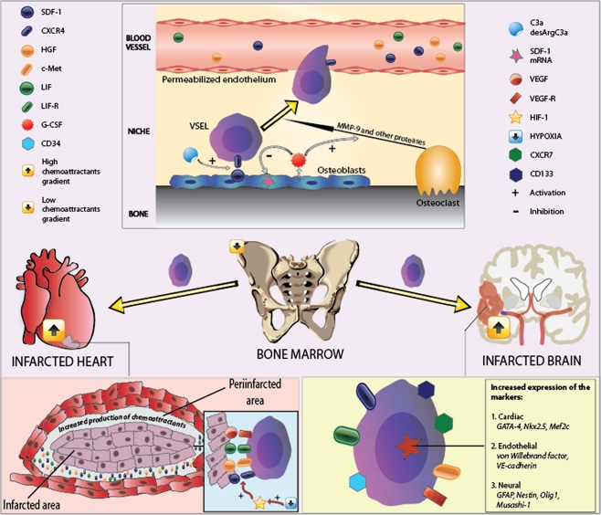
Very small embryonic-like cells (VSELs) are a population of stem cells residing in the bone marrow (BM) and several organs, which undergo mobilization into peripheral blood (PB) following acute myocardial infarction and stroke. These cells express markers of pluripotent stem cells (PSCs), such as Oct-4, Nanog, and SSEA-1, as well as early cardiac, endothelial, and neural tissue developmental markers. VSELs can be effectively isolated from the BM, umbilical cord blood, and PB. Peripheral blood and BM-derived VSELs can be expanded in co-culture with C2C12 myoblast feeder layer and undergo differentiation into cells from all three germ layers, including cardiomyocytes and vascular endothelial cells. Isolation of VSLEs using fluorescence-activated cell sorting multiparameter live cell sorting system is dependent on gating strategy based on their small size and expression of PSC and absence of hematopoietic lineage markers. VSELs express early cardiac and endothelial lineages markers (GATA-4, Nkx2.5/Csx, VE-cadherin, and von Willebrand factor), SDF-1 chemokine receptor CXCR4, and undergo rapid mobilization in acute MI and ischemic stroke. Experiments in mice showed differentiation of BM-derived VSELs into cardiac myocytes and effectiveness of expanded and pre-differentiated VSLEs in improvement of left ventricular ejection fraction after myocardial infarction.


Figures

Fig. 1
Fig. 1
Strategy for isolation of VSELs from human peripheral blood after mobilization with C-CSF (MPB-VSELs) using FACS-based live cell sorting system. Gating strategy was developed by using of the synthetic beads of known diameters (1, 2, 4, 6, 10, 15 μm) (a) to define the extended lymph-gate for subsequent sorting (b). After lysis of erythrocytes, mobilized peripheral blood total nucleated cells (TNCs) fraction is stained with antibodies against hematopoietic lineages markers (Lin), CD133 stem cell antigen (c), and CD45 pan-leukocytic antigen (d). Gate R1 was set up to include objects with diameter > 2 μm. Events included in region R1 were analyzed for presence of hematopoietic lineages markers. Subsequently, only lin events were included into region R2, and cells expressing CD133 antigens were further isolated depending on the presence of CD45 antigen (gates R3-4). VSELs are linCD45-CD133+ cells (gate R3) whereas hematopoietic stem cells (HPCs) are included in gate R4 and constitute population of linCD45+CD133+
Fig. 2
Fig. 2
Representative images of human peripheral blood-derived VSEL and HSPC by ImageStreamX system. Human blood cells were stained for markers distinguishing VSELs such as: (1) CD45 pan-leukocytic antigen (APC-Cy7, cyan), (2) hematopoietic lineages markers (FITC, green) and (3) stem cell antigens CD133 (PE, yellow), and CD34 (APC, violet). Nuclei were stained with Hoechst 33342 dye. Images were collected by imaging flow cytometer-ImageStreamX system. VSELs and HSPCs were distinguished based on CD45 antigen expression
Fig. 3
Fig. 3
The putative mechanism of mobilization and homing of VSELs in acute myocardial infarction. Myocardial ischemia induces increased expression of chemoattractants [chemokines (SDF-1), growth factors (VEGF, HGF), cytokines (LIF)] and release of phospholipids predominantly in infarct border zone. The increased expression of chemoattractants in the ischemic organ creates the reversal of chemoattractant gradient leading to the release of VSELs from the bone marrow niches and their homing to the site of the ischemic injury. Within the bone marrow niche, the mobilization of VSELs is orchestrated by expression of matrix metaloproteinases and activation of complement cascade. Also, phospholipids, such as sphingosine-1-phosphate influence the mobilization of VSELs. In peripheral blood in healthy subjects, VSELs can be detected at a very low number. After ischemic injury, these cells are rapidly mobilized into peripheral blood and the expression of pluripotent stem cell markers, as well as early cardiac, endothelial, muscle, and neural markers is significantly increased. Hence, we hypothesized that mobilization of VSELs is a part of reparatory mechanism activated in the setting of acute myocardial infarction. Also, circulation of VSELs might contribute to the pool of resident cardiac stem cells

References

    1. Abdel-Latif A, Bolli R, Tleyjeh IM, Montori VM, Perin EC, Hornung CA, et al. Adult bone marrow-derived cells for cardiac repair: a systematic review and meta-analysis. Archives of Internal Medicine. 2007;167(10):989–997. doi: 10.1001/archinte.167.10.989. - DOI - PubMed
    1. Dimmeler S, Burchfield J, Zeiher AM. Cell-based therapy of myocardial infarction. Arteriosclerosis, Thrombosis, and Vascular Biology. 2008;28(2):208–216. doi: 10.1161/ATVBAHA.107.155317. - DOI - PubMed
    1. Messina E, De Angelis L, Frati G, Morrone S, Chimenti S, Fiordaliso F, et al. Isolation and expansion of adult cardiac stem cells from human and murine heart. Circulation Research. 2004;95(9):911–921. doi: 10.1161/01.RES.0000147315.71699.51. - DOI - PubMed
    1. Behfar, A., Yamada, S., Crespo-Diaz, R., Nesbitt, J. J., Rowe, L. A., Perez-Terzic, C., et al. Guided cardiopoiesis enhances therapeutic benefit of bone marrow human mesenchymal stem cells in chronic myocardial infarction. Journal of the American College of Cardiology, 569, 721–734. - PMC - PubMed
    1. Pittenger MF, Martin BJ. Mesenchymal stem cells and their potential as cardiac therapeutics. Circulation Research. 2004;95(1):9–20. doi: 10.1161/01.RES.0000135902.99383.6f. - DOI - PubMed

Publication types

MeSH terms