. 2011 Jan;79(1):21-34.
doi: 10.1111/j.1365-2958.2010.07439.x. Epub 2010 Nov 16.

Association of OLE RNA with bacterial membranes via an RNA-protein interaction

Affiliations

Association of OLE RNA with bacterial membranes via an RNA-protein interaction

Kirsten F Block et al. Mol Microbiol. 2011 Jan.

Abstract

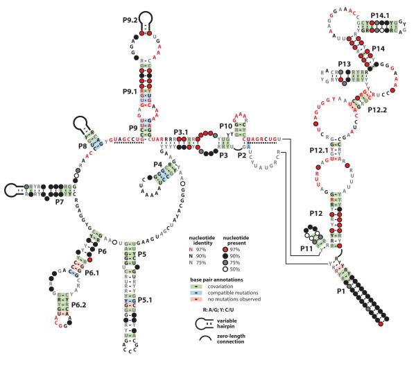
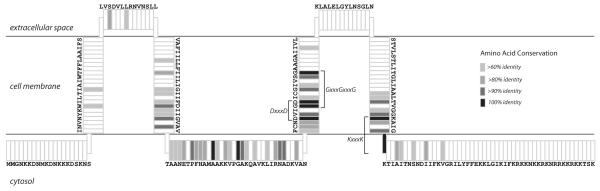
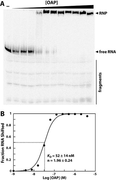
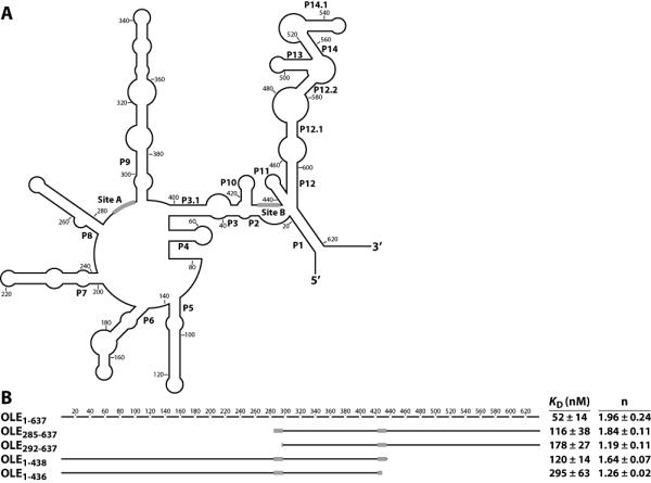
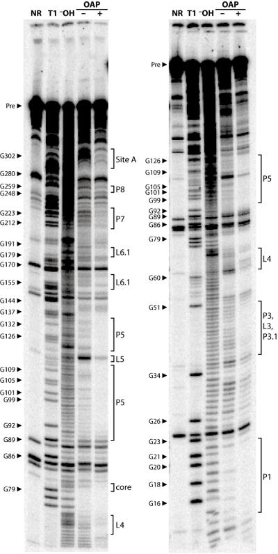
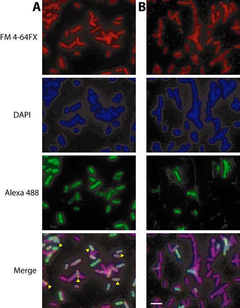
Ornate, large, extremophilic (OLE) RNAs are large, non-coding transcripts characterized by their ornate secondary structure and presence predominantly in Gram-positive, extremophilic bacteria. A gene for an OLE-associated protein (OAP) is almost always located immediately downstream of the OLE gene. OAP has no extensive homology to other proteins and is predicted to form multiple transmembrane domains. We show that this protein forms a ribonucleoprotein complex with OLE RNA using at least 2:1 protein : RNA stoichiometry. A series of truncated OLE RNA constructs was used to establish that most of the RNA can be deleted without eliminating protein binding. Two primary binding sites are present within the RNA, although additional binding determinants exist and extensive structural stabilization is induced by OAP. RNA fluorescence in situ hybridization (FISH) was used in Escherichia coli to demonstrate that ribonucleoprotein complex formation localizes the RNA near cell membranes of this heterologous system. Therefore, the majority of the complex structure formed by OLE RNA may perform a biochemical function that requires membrane localization.


Figures

Fig. 1
Fig. 1
Revised consensus sequence and secondary structure model for OLE RNAs. Phylogenetic analysis of 78 examples from sequenced genomes, metagenomic data, and community PCR were used to define the consensus model. Dashed lines indicate highly conserved regions implicated in protein binding.
Fig. 2
Fig. 2
Transcriptome deep sequencing reveals that OLE RNA is highly expressed in B. halodurans. (A) Listed are the fifteen most common RNAs in B. halodurans C-125 other than ribosomal and transfer RNAs. Both rRNAs and tRNAs are likely more abundant than those RNAs listed but were eliminated from the RNA sample before cDNA generation. Relative abundance was determined by calculating RPKM scores (Reads Per Kilobase per Million reads (Mortazavi et al., 2008)) for each RNA before normalizing to OLE RNA. (B) The average reads per nucleotide using a 50-base-pair window are graphed for the genomic region (bottom of graph) corresponding to the OLE RNA-containing operon as defined previously (Puerta-Fernandez et al., 2006) (see Table S1 for raw data).
Fig. 3
Fig. 3
Consensus and topology of OLE-Associated Proteins. The consensus sequence and predicted topology of OAP are shown with transmembrane domains depicted as vertical segments and extracellular or cytosolic domains shown as horizontal segments. Orientation with respect to the cytosol and extracellular space is indicated. Shading depicts conservation of primary sequence, with black positions representing 100% amino acid conservation across all known examples.
Fig. 4
Fig. 4
OLE and OAP form a ribonucleoprotein complex. (A) 5′ 32P-labeled OLE1-637 RNA was subjected to electrophoretic mobility shift assays with increasing concentrations of N-terminal histidine-tagged OAP to assess ribonucleoprotein complex formation. Arrowheads denote free, full-length RNA and shifted RNA (RNP) as noted. A bar highlights residual 3′ degradation products (fragments). These degraded RNAs do not shift in mobility in response to OAP. (B) To determine the binding characteristics of OAP, the fraction OLE RNA shifted was plotted versus the logarithm of the concentration of OAP. The data depicted is a representative and plots data calculated from the gel shown in part A. The KD and Hill coefficient (n) were calculated from four separate experiments, and the average and standard error are listed on the graph.
Fig. 5
Fig. 5
Identification of nucleotides necessary for formation of the OLE-OAP complex. (A) Secondary structure of the B. halodurans OLE RNA sequence is depicted in outline. Black bars indicate the predicted OAP binding sites as determined in B. (B) Full-length OLE RNA is depicted as a broken line, with each line segment corresponding to 20 nucleotides. Solid lines correspond to the indicated truncated constructs, with approximate location depicted graphically. The KD and Hill coefficient for each construct were calculated from at least four experiments and are given with the standard error. From these truncations, predicted OAP-binding sites were identified and depicted as black boxes on both the truncated RNAs and in part A. No binding (n.b.) was observed for the construct OLE285-438.
Fig. 6
Fig. 6
Probing of OLE RNA with OAP reveals structural modulation in response to protein binding. 5′ 32P-labeled OLE1-637 RNA was subjected to Pb2+ -catalyzed cleavage in the presence (+) or absence (□) of OAP to identify regions of OAP-mediated structural modulation. Other lane assignments are as follows: NR, no reaction; T1, partial digestion with RNase T1 under denaturing conditions; OH, partial digestion with alkali. Select bands corresponding to RNase T1 digestion products (cleavage after G residues) are labeled. The locations of structural elements as defined in Fig. 1 are indicated. Bands in the no reaction lanes are fragments produced by spontaneous cleavage during the long elution times needed to recover precursor (Pre) RNAs from purification gels.
Fig. 7
Fig. 7
Subcellular localization of exogenously expressed OLE RNA when co-expressed with OAP (A) or expressed alone (B). E. coli cells expressing OLE with or without OAP were probed for cellular localization of OLE RNA using fluorescence in situ hybridization (RNA FISH). Fluorescence signal from the membrane stain (FM 4-64 FX, top), nucleic acids (DAPI, second), antisense RNA probe (Alexa 488, third) and merge (bottom) are shown. Yellow arrowheads indicate cells with overlap between RNA localization and the membrane stain. The scale bar in the lower right panel denotes 5 μm.

Comment in

  • Membrane RNAs in bacteria.
    Sanders W, Laederach A. Sanders W, et al. Mol Microbiol. 2011 Jan;79(1):1-2. doi: 10.1111/j.1365-2958.2010.07438.x. Mol Microbiol. 2011. PMID: 21210530 Free PMC article. No abstract available.

References

    1. Baliarda A, Robert H, Jebbar M, Blanco C, Le Marrec C. Isolation and characterization of ButA, a secondary glycine betaine transport system operating in Tetragenococcus halophila. Curr Microbiol. 2003;47:347–351. - PubMed
    1. Bartel DP, Chen CZ. Micromanagers of gene expression: the potentially widespread influence of metazoan microRNAs. Nat Rev Genet. 2004;5:396–400. - PubMed
    1. Block KF, Hammond MC, Breaker RR. Evidence for widespread gene control function by the ydaO riboswitch candidate. J Bacteriol. 2010;192:3983–3989. - PMC - PubMed
    1. Brandt W, Brauer L, Gunnewich N, Kufka J, Rausch F, Schulze D, et al. Molecular and structural basis of metabolic diversity mediated by prenyldiphosphate converting enzymes. Phytochemistry. 2009;70:1758–1775. - PubMed
    1. Corbino KA, Barrick JE, Lim J, Welz R, Tucker BJ, Puskarz I, et al. Evidence for a second class of S-adenosylmethionine riboswitches and other regulatory RNA motifs in alpha-proteobacteria. Genome Biol. 2005;6 - PMC - PubMed

MeSH terms