. 2010 Dec 18:8:32.
doi: 10.1186/1477-3155-8-32.

Manufacturing and in vivo inner ear visualization of MRI traceable liposome nanoparticles encapsulating gadolinium

Affiliations

Manufacturing and in vivo inner ear visualization of MRI traceable liposome nanoparticles encapsulating gadolinium

Jing Zou et al. J Nanobiotechnology. .

Abstract

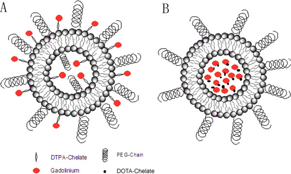
Background: Treatment of inner ear diseases remains a problem because of limited passage through the blood-inner ear barriers and lack of control with the delivery of treatment agents by intravenous or oral administration. As a minimally-invasive approach, intratympanic delivery of multifunctional nanoparticles (MFNPs) carrying genes or drugs to the inner ear is a future therapy for treating inner ear diseases, including sensorineural hearing loss (SNHL) and Meniere's disease. In an attempt to track the dynamics and distribution of nanoparticles in vivo, here we describe manufacturing MRI traceable liposome nanoparticles by encapsulating gadolinium-tetra-azacyclo-dodecane-tetra-acetic acid (Gd-DOTA) (abbreviated as LPS+Gd-DOTA) and their distribution in the inner ear after either intratympanic or intracochlear administration.

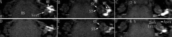
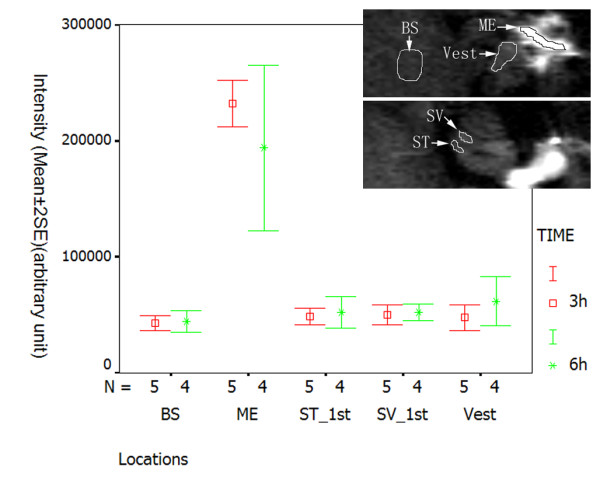
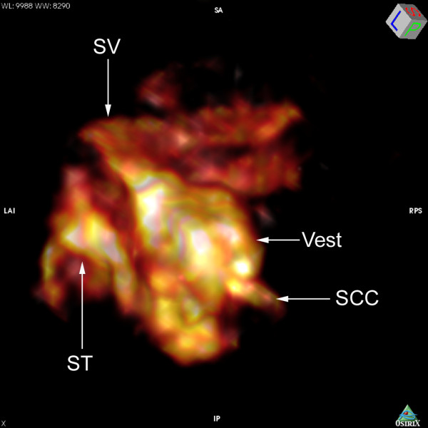
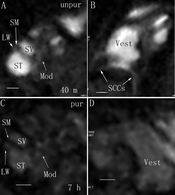
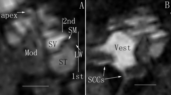
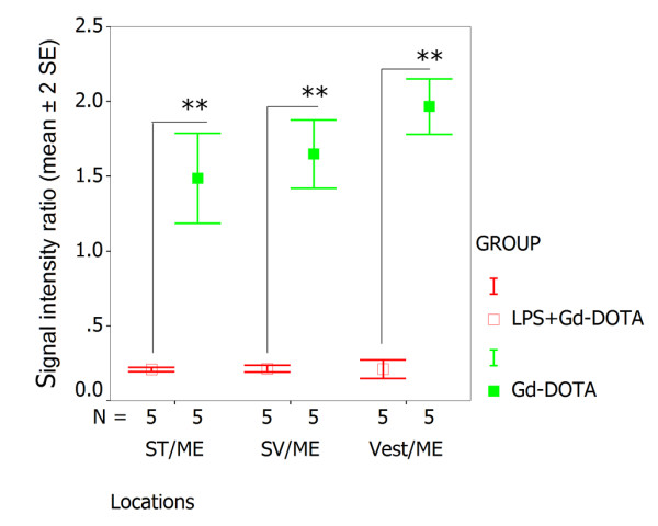
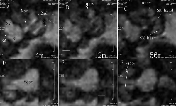
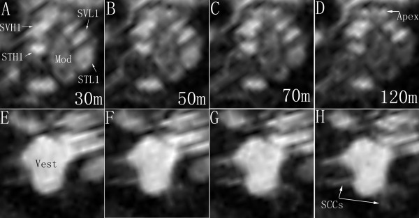
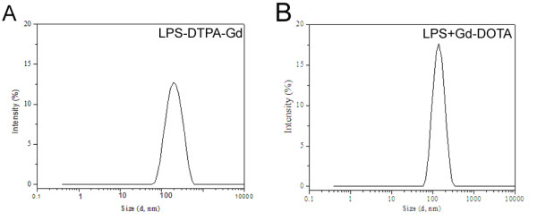
Results: Measurements of relaxivities (r1 and r2) showed that LPS+Gd-DOTA had efficient visible signal characteristics for MRI. In vivo studies demonstrated that LPS+Gd-DOTA with 130 nm size were efficiently taken up by the inner ear at 3 h after transtympanic injection and disappeared after 24 h. With intracochlear injection, LPS+Gd-DOTA were visualized to distribute throughout the inner ear, including the cochlea and vestibule with fast dynamics depending on the status of the perilymph circulation.

Conclusion: Novel LPS+Gd-DOTA were visible by MRI in the inner ear in vivo demonstrating transport from the middle ear to the inner ear and with dynamics that correlated to the status of the perilymph circulation.


Figures

Figure 1
Figure 1
Illustration of rat inner ear anatomy and transportation routes of substances to be gained in the inner ear after different administrations. After intratympanic delivery, agents may enter the inner ear through the round window membrane (1) and annular ligament (2), which surrounds the stapes footplate within the oval window and comprises the stapediovestibular joint (3). Stapedial artery (4) is a prominant structure in the rat middle ear, which runs through the obturator foramen of the stapes and inferior to the round window niche. The inner ear is composed of the cochlea (5) and vestibule. The vestibule contains three semicircular canals including the lateral (6), posterior (7) and superior canals (8), and the saccule (9) and utricle (10). The crista (11), which is located in each ampulla, is the sensory structure of the semicircular canals. The macula (12) is the sensory structure of both the saccule and utricle. The cochlea contains three chambers, which are the scala tympani (13), the scala vestibuli (14), and the scala media (15). The scala tympani and vestibuli are filled by perilymph (high sodium-containing fluid). The scala media is filled with endolymph (high potassium-containing fluid) and also contains the sensory structure, the organ of Corti (16). The basilar membrane (17), which extends from the osseous spiral lamina (18), separates the scala media from scala tympani. Reissner's membrane (19) separates the scala media from scala vestibuli. Spiral ganglion cells (20), which are located in the modiolus, send peripheral processes to the hair cells and central processes to the cochlear nuclei via cochlear nerve. Capillaries in the modiolus (21) and spiral ligament (22) contribute to the blood-perilymph barrier, which is the transportation pathway of agents to reach the inner ear after intravenous or oral administration. Capillaries in the stria vascularis (23) are attributable to the blood-endolymph barrier.
Figure 2
Figure 2
Visualization of LPS+Gd-DOTA in the middle ear cavity and inner ear of rat after transtympanic injection using T1-weighted 2D MRI. LPS+Gd-DOTA nanoparticles (1.0 mM LPS+Gd-DOTA containing 500 mM Gd-DOTA) were delivered to the left middle ear. At 3 h time point (A-C), they were detected in the middle ear cavity, vestibule, and cochlea. Aftert 6 h (D-F), these regions with uptake of nanoparticles showed brighter signal and the second turn and apex of the cochlea became more visible. 1st: the basal turn; 2nd: the second turn; BS: brainstem; L: left ear; ME: middle ear; R: right ear; ST: scala tympani; SV: scala vestibuli; Vest: vestibule. Particle sizes = 150 ± 20 nm. Scale bar = 1 mm.
Figure 3
Figure 3
Semi-quantification of LPS+Gd-DOTA in the middle ear cavity and inner ear of rat after transtympanic injection demonstrated by T1-weighted 2D MRI. 1.0 mM LPS+Gd-DOTA (containing 500 mM Gd-DOTA) was administered. At 3 h post-administration, LPS+Gd-DOTA nanoparticles were efficiently detected in the perilymph, which reached 21.1% (average of the vestibule, scala vestibuli, and scala tympani) of that in the middle ear cavity. LPS+Gd-DOTA intensity decreased in the middle ear cavity while increased in the inner ear and brainstem form 3 h to 6 h after intra tympanic administration. The region of interest was shown in the images. BS: brainstem; ME: middle ear; ST: scala tympani; SV: scala vestibuli; Vest: vestibule.
Figure 4
Figure 4
3D rendering of T1-weighted MRI demonstrating spatial distribution of LPS+Gd-DOTA in the rat inner ear after transtympanic injection. 1.0 mM LPS+Gd-DOTA (containing 500 mM Gd-DOTA) was administered. Abundant nanoparticles retained in the vestibule (Vest) and perilymphatic compartments of the basal lower turn. SCC: semicircular canal; ST: scala tympani; SV: scala vestibuli.
Figure 5
Figure 5
Different uptake of unpurified and purified LPS+Gd-DOTA in rat inner ear after transtympanic injection shown by MPR single view of T1-weighted 3D MR imaging. Unpurified LPS+Gd-DOTA were efficiently taken up in the inner ear at 40 min after transtympanic injection at a concentration of 1 mM (containing 500 mM Gd-DOTA) (A and B). The signal intensity of purified LPS+Gd-DOTA (B and C) in the inner ear was significantly lower than that of unpurified LPS+Gd-DOTA judged by vision. LW: lateral wall; Mod: modiolus; MPR: multiplanar reconstruction; SM: scala media; ST: scala tympani; SV: scala vestibuli; Vest: vestibule; SCCs: semicircular canals; unpur: unpurified LPS+Gd-DOTA; pur: purified LPS+Gd-DOTA. Particle sizes = 300 ± 20 nm. Sacle bar = 500 μm.
Figure 6
Figure 6
MPR single view of T1-weighted 3D MR imaging of the rat inner ear after transtympanic injection of Gd-DOTA. Gd-DOTA distributed universally in the cochlea and vestibule at 3 h post-transtympanic injection. LW: lateral wall; Mod: modiolus; MPR: multiplanar reconstruction; SCCs: semicircular canals; SM: scala media; ST: scala tympani; SV: scala vestibuli; Vest: vestibule. Scale bar = 1 mm.
Figure 7
Figure 7
Semi-quantitative comparison of signal ratios in the perilymphatic compartments over the middle ear cavity between LPS+Gd-DOTA and Gd-DOTA acquired by T1-weighted 2D MRI. LPS+Gd-DOTA was transported less effectively than Gd-DOTA alone through the middle-inner ear barriers (**p < 0.01, student t-test). ME: middle ear; ST: scala tympani; SV: scala vestibuli; Vest: vestibule.
Figure 8
Figure 8
Dynamic distribution of LPS+Gd-DOTA in the inner ear after intracochlear delivery in closed perilymphatic compartments demonstrated by MPR single view of T1-weighted 3D MRI. 1 mM nanoparticles containing 500 mM Gd-DOTA were administered. In the cochlea, LPSG+Gd-DOTA filled SM at the beginning, but were cleaned from SM after 56 m (A, B, and C). In the vestibule, LPS+Gd-DOTA retained in the proximal locations of SCCs, but distributed to the distal parts of SCCs at 56 m post-injection (D, E, F). 1st: the basal turn; 2nd:the second turn; 4 m, 12 m, and 56 m: 4 minutes, 12 minutes, and 56 minutes; Mod: modiolus; MPR: multiplanar reconstruction; SSCs: semicircular canals; SM: the scala media; SM-h1st: SM of the higher basal turn; SM-h2nd: SM of the higher second turn; ST: the scala tympani; SV: the scala vestibuli; Vest: vestibule. Scale bar = 500 μm.
Figure 9
Figure 9
Dynamic distribution of LPS+Gd-DOTA in the inner ear after intracochlear delivery in an open perilymphatic compartments demonstrated by MPR single view of T1-weighted 3D MRI. 1 mM nanoparticle containing 500 mM Gd-DOTA were administered. Intracochlear dynamics was shown in A through D; intravestabular dynamics was shown in E through H. From 30 min to 120 min post-administration of LPS+Gd-DOTA, signal intensities decayed gradually in the STL1, STH1, and Mod; but retained stable in the SVL1, SVH1, and Vest. 30 m, 50 m, etc.: 30 minutes etc; Mod: modilous; MPR: multiplanar reconstruction; SSCs: semicircular canals; STH1: scala tympani of the higher basal turn; STL1: scala tympani of the lower basal turn; SVH1: scala vestibuli of the higher basal turn; SVL1: scala vestibuli of the lower basal turn; Vest: vestibule.
Figure 10
Figure 10
Schematic representation of liposomes containing gadolinium chelate lipid (LPS-DTPA-Gd) (and liposomes encapsulating Gd-DOTA (LPS+Gd-DOTA). A: LPS-DTPA-Gd; B: LPS+Gd-DOTA.
Figure 11
Figure 11
Size distribution by intensity of purified liposomes (total lipid concentration 1 mM) containing DTPA-Gd (LPS-DTPA-Gd) or encapsulating Gd-DOTA (LPS+Gd-DOTA) obtained by dynamic light scattering. Temperature was maintained at 25°C. LPS-DTPA-Gd (A) showed more dispersed size distribution than LPS+Gd-DOTA (B).

References

    1. Zou J, Zhang W, Poe D, Qin J, Fornara A, Zhang Y, Ramadan UA, Muhammed M, Pyykko I. MRI manifestation of novel superparamagnetic iron oxide nanoparticles in the rat inner ear. Nanomedicine (Lond) pp. 739–754. - DOI - PubMed
    1. Nanoear: 3g-Nanotechnology based targeted drug delivery using the inner ear as a model target organ. http://www.nanoear.org/
    1. Zou J, Saulnier P, Perrier T, Zhang Y, Manninen T, Toppila E, Pyykko I. Distribution of lipid nanocapsules in different cochlear cell populations after round window membrane permeation. J Biomed Mater Res B Appl Biomater. 2008;87:10–18. - PubMed
    1. Zou J, Zhang Y, Zhang W, Ranjan S, Sood R, Mikhailov A, Kinnunen P, Pyykko I. Internalization of liposome nanoparticles functionalized with TrkB ligand in rat cochlear cell populations. European Journal of Nenomedicine. 2009;3:8–14.
    1. Ishii T. The fine structure of lipofuscin in the human inner ear. Arch Otorhinolaryngol. 1977;215:213–221. doi: 10.1007/BF00463059. - DOI - PubMed