. 2011 Feb 20;410(2):291-8.
doi: 10.1016/j.virol.2010.11.016. Epub 2010 Dec 17.

XPB mediated retroviral cDNA degradation coincides with entry to the nucleus

Affiliations

XPB mediated retroviral cDNA degradation coincides with entry to the nucleus

Kristine E Yoder et al. Virology. .

Abstract

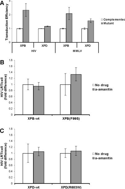
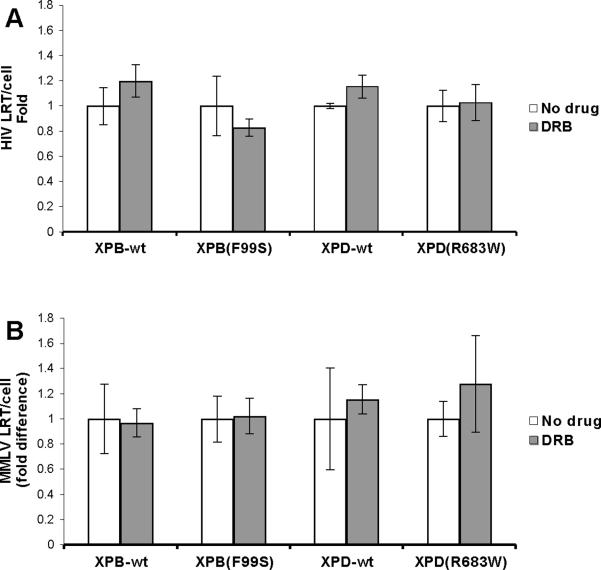
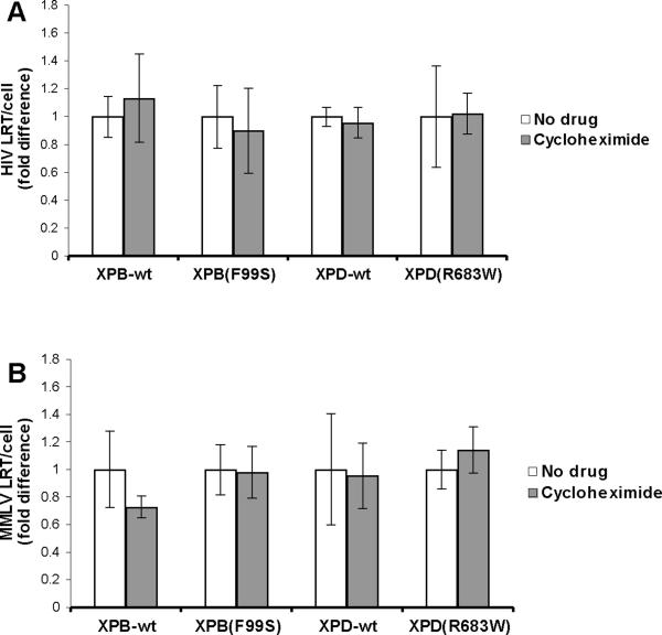
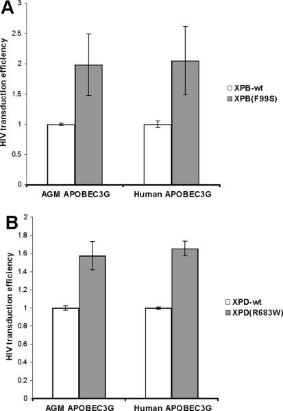
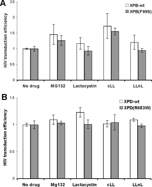
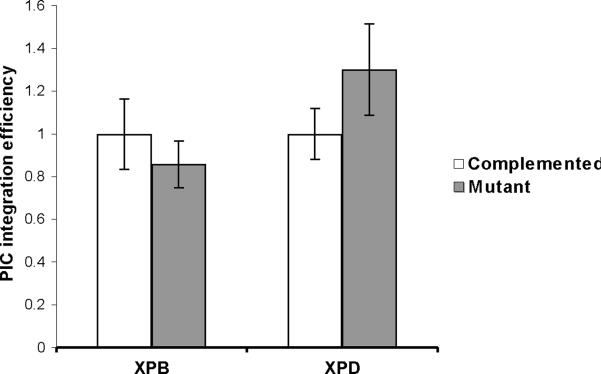
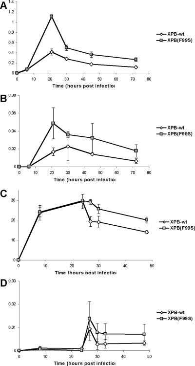
Retroviruses must integrate their cDNA to a host chromosome, but a significant fraction of retroviral cDNA is degraded before integration. XPB and XPD are part of the TFIIH complex which mediates basal transcription and DNA nucleotide excision repair. Retroviral infection increases when XPB or XPD are mutant. Here we show that inhibition of mRNA or protein synthesis does not affect HIV cDNA accumulation suggesting that TFIIH transcription activity is not required for degradation. Other host factors implicated in the stability of cDNA are not components of the XPB and XPD degradation pathway. Although an increase of retroviral cDNA in XPB or XPD mutant cells correlates with an increase of integrated provirus, the integration efficiency of pre-integration complexes is unaffected. Finally, HIV and MMLV cDNA degradation appears to coincide with nuclear import. These results suggest that TFIIH mediated cDNA degradation is a nuclear host defense against retroviral infection.


Figures

Fig. 1
Fig. 1
Retroviral infection of wild type and NER mutant cell lines. (A) An equal number of NER mutant (XPB(F99S) and XPD(R683W)) and complemented cells (XPB-wt and XPD-wt) were infected with HIV or MMLV retroviral vectors expressing GFP following integration to the host genome. At 72 hpi the cells were analyzed by flow cytometry for GFP expression. Transduction efficiency is expressed relative to the complemented cells expressing the wild type gene. (B and C) HIV late reverse transcripts in wild type and NER mutant cells treated with the transcription inhibitor α-amanitin. An equal number of wild type and NER defective cells were treated with the RNA polymerase II inhibitor α-amanitin for 2 hours and infected with an HIV based retroviral vector in the continued presence of α-amanitin (a-amanitin). DNA was collected at 6 hours after the addition of HIV. HIV late reverse transcripts (LRT) and the cellular 18S gene were measured by qPCR. The quantity of HIV LRT was divided by the number of cellular genomes to obtain the number of HIV LRT per cell (HIV LRT/cell). (B) XPB-wt and NER defective XPB(F99S) cell lines. (C) XPD-wt and NER defective XPD(R683W) cell lines. Data from α-amanitin treated cells is expressed relative to untreated cells (No drug). Error bars indicate the standard deviation between triplicates at two MOI in three independent experiments.
Fig. 2
Fig. 2
HIV late reverse transcripts in XP cell lines treated with a CAK inhibitor. Wild type and NER mutant cells were treated with the CAK inhibitor DRB for 2 hours and infected with an HIV based retroviral vector in the continued presence of the drug. DNA was collected at 6 hours after the addition of HIV. HIV late reverse transcripts (LRT) and the cellular 18S gene were measured by qPCR and used to determine HIV LRT per cell. (A) XPB-wt and NER defective XPB(F99S) cell lines. (B) XPD-wt and NER defective XPD(R683W) cell lines. Data from DRB treated cells is expressed relative to untreated cells (No drug). Error bars indicate the standard deviation between triplicates at two MOI in three independent experiments.
Fig. 3
Fig. 3
HIV late reverse transcripts in wild type and NER mutant XP cell lines treated with the translation inhibitor cycloheximide. An equal number of (A) XPB-wt and XPB(F99S) or (B) XPD-wt and XPD(R683W) cells were treated with the ribosome inhibitor cycloheximide for 2 hours and infected with an HIV based retroviral vector in the continued presence of cycloheximide. DNA was collected at 6 hours after the addition of HIV particles. HIV late reverse transcripts (LRT) and the cellular 18S gene were measured by qPCR. The quantity of HIV LRT was divided by the number of cellular genomes to obtain the number of HIV LRT per cell (HIV LRT/cell). Data from cycloheximide treated cells is expressed relative to untreated cells (No drug). Error bars indicate the standard deviation between triplicates at two MOI in three independent experiments.
Fig. 4
Fig. 4
Role of TFIIH in the APOBEC3G host defense pathway. HIV vector particles including Vif were generated in the presence of African green monkey (AGM) or human APOBEC3G. (A) XPB-wt and XPB(F99S) or (B) XPD-wt and XPD(R683W) cells were infected with AGM or human APOBEC3G HIV vectors. The HIV vector expresses GFP following integration. Cells were assayed by flow cytometry for GFP expression from the integrated HIV cDNA. The infection efficiency of mutant cell lines is expressed relative to wild type cells and % difference graphed. Error bars indicate the standard deviation between duplicates at two MOI in two independent experiments.
Fig. 5
Fig. 5
Effects of proteasome inhibitors on HIV infection efficiency in XP cell lines. (A) XPB-wt and XPB(F99S) cells were infected with HIV vector particles in the presence of 2 μM MG132, 2 μM lactacystin, 2 μM clasto-lactacystin β-lactone (cLL), or 5 μM calpain inhibitor I (LLnL). (B) XPD-wt and XPD(R683W) cells were infected with HIV vector particles in the presence of 0.5 μM MG132, 2.5 μM lactacystin, 1 μM clasto-lactacystin β-lactone (cLL), or 50 μM calpain inhibitor I (LLnL). The HIV vector particles express GFP following integration and cells were assayed by flow cytometry for GFP expression 72hpi. Infection efficiency is expressed relative to cells infected in the presence of DMSO (No drug). Error bars indicate the standard deviation between duplicates at two MOI in three independent experiments.
Fig. 6
Fig. 6
Integration efficiency of PICs derived from XP cell lines. XPB-wt (complemented), XPB(F99S), XPD-wt (complemented), and XPD(R683W) cell lines were infected with HIV vector particles. PIC extracts were harvested at 6 hpi. 100 ng human genomic DNA was added to PICs as an integration target and incubated at 37°C for 1 hour. DNA was purified and assayed by qPCR for late reverse transcripts and integration products. The PIC integration efficiency is expressed relative to PICs from wild type complemented cells. Error bars indicate the standard deviation between duplicate integration reactions from two independent preparations of PICs.
Fig. 7
Fig. 7
Infection of XPB cell lines in the presence of aphidicolin. XPB-wt (open diamonds) and XPB(F99S) (filled squares) cells were treated with 1 μg/ml aphidicolin for 18 hours before infection and arrested at G1/S. HIV vector particles were added in the continued presence of aphidicolin. DNA was collected at 6, 24, 30, 48, and 72 hours post infection and analyzed by qPCR for the accumulation of (A) HIV late reverse transcripts (LRT), (B) HIV 2LTR circles. The 18S gene was quantified to yield the number of HIV cDNAs per cell. (C and D) XPB-wt (open diamonds) and XPB(F99S) (filled squares) cells were treated with 1 μg/ml aphidicolin for 18 hours before infection and arrested at G1/S. MMLV vector particles were added in the continued presence of aphidicolin. Aphidicolin was removed at 24 hpi. DNA was collected at 8, 24, 27, 30, and 48 hours post infection and analyzed by qPCR for the accumulation of (C) MMLV late reverse transcripts (LRT) and (D) MMLV 2LTR circles. The 18S gene was quantified to yield the number of MMLV LRT per cell. Error bars indicate the standard deviation between duplicates in two independent experiments.

References

    1. Albin JS, Harris RS. Interactions of host APOBEC3 restriction factors with HIV-1 in vivo: implications for therapeutics. Expert Rev Mol Med. 2010;12:e4. - PMC - PubMed
    1. Barbosa P, Charneau P, Dumey N, Clavel F. Kinetic analysis of HIV-1 early replicative steps in a coculture system. AIDS Res Hum Retroviruses. 1994;10(1):53–9. - PubMed
    1. Benhamou S, Sarasin A. ERCC2 /XPD gene polymorphisms and lung cancer: a HuGE review. Am J Epidemiol. 2005;161(1):1–14. - PubMed
    1. Biglione S, Byers SA, Price JP, Nguyen VT, Bensaude O, Price DH, Maury W. Inhibition of HIV-1 replication by P-TEFb inhibitors DRB, seliciclib and flavopiridol correlates with release of free P-TEFb from the large, inactive form of the complex. Retrovirology. 2007;4:47. - PMC - PubMed
    1. Bishop KN, Holmes RK, Malim MH. Antiviral potency of APOBEC proteins does not correlate with cytidine deamination. J Virol. 2006;80(17):8450–8. - PMC - PubMed

Publication types

MeSH terms