. 2011 Mar;32(7):1890-905.
doi: 10.1016/j.biomaterials.2010.11.028. Epub 2010 Dec 16.

Multi-functional magnetic nanoparticles for magnetic resonance imaging and cancer therapy

Affiliations

Multi-functional magnetic nanoparticles for magnetic resonance imaging and cancer therapy

Murali M Yallapu et al. Biomaterials. 2011 Mar.

Abstract

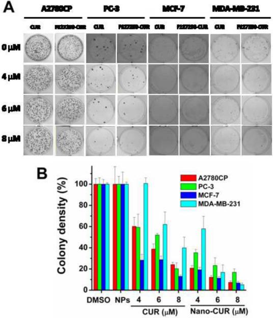
We have developed a multi-layer approach for the synthesis of water-dispersible superparamagnetic iron oxide nanoparticles for hyperthermia, magnetic resonance imaging (MRI) and drug delivery applications. In this approach, iron oxide core nanoparticles were obtained by precipitation of iron salts in the presence of ammonia and provided β-cyclodextrin and pluronic polymer (F127) coatings. This formulation (F127250) was highly water dispersible which allowed encapsulation of the anti-cancer drug(s) in β-cyclodextrin and pluronic polymer for sustained drug release. The F127250 formulation has exhibited superior hyperthermia effects over time under alternating magnetic field compared to pure magnetic nanoparticles (MNP) and β-cyclodextrin coated nanoparticles (CD200). Additionally, the improved MRI characteristics were also observed for the F127250 formulation in agar gel and in cisplatin resistant ovarian cancer cells (A12780CP) compared to MNP and CD200 formulations. Furthermore, the drug-loaded formulation of F127250 exhibited many folds of imaging contrast properties. Due to the internalization capacity of the F127250 formulation, its curcumin-loaded formulation (F127250-CUR) exhibited almost equivalent inhibition effects on A2780CP (ovarian), MDA-MB-231 (breast), and PC-3 (prostate) cancer cells even though curcumin release was only 40%. The improved therapeutic effects were verified by examining molecular effects using Western blotting and transmission electron microscopic (TEM) studies. F127250-CUR also exhibited haemocompatibility, suggesting a nanochemo-therapeutic agent for cancer therapy.


Figures

Fig. 1
Fig. 1
Particles size characterization of magnetic nanoparticle formulations: (A) Dynamic light scattering particles size data of (a) pure magnetic nanoparticles (MNP), (b) magnetic nanoparticles coated with 200 mg of CD (CD200) and (c) magnetic nanoparticles coated with 200 mg of CD and 250 mg of F127 polymer (F127250). (B) Transmission electron microscopic images of (a) pure magnetic nanoparticles, (b) magnetic nanoparticles coated with 200 mg of CD and (c) magnetic nanoparticles coated with 200 mg of CD and 250 mg of F127 polymer. (C) Transmission electron microscopic image of (a) pure magnetic nanoparticles, (b) magnetic nanoparticles coated with 200 mg of CD and (c) magnetic nanoparticles coated with 200 mg of CD and 250 mg of F127 polymer. Data showing individual particle grain size of 7–10 nm.
Fig. 2
Fig. 2
Physical characterization of magnetic nanoparticle formulations: (A) X-ray diffraction patterns, (B) Fourier transform infrared spectra, and (C) thermograms of MNP, CD200, and F127250 nanoparticle formulations. (D) Curcumin release profiles from curcumin loaded MNP, CD200, and F127250 formulations. Cumulative release was estimated using UV-vis spectrophotometric method. Data presented is a mean of three replicates. Note: (B) also presents curcumin and curcumin containing F127250 nanoparticle formulations (F127250-CUR).
Fig. 3
Fig. 3
(A) Hysteresis loops of MNP, CD200 and F127250 nanoparticle formulations at room temperature. (B) Time course of the raised temperature of MNP, CD200 and F127250 nanoparticle formulations under an alternating magnetic field operating at 300 kHz. (C) Temperature of various concentrations of F127250 nanoparticles in solution and agarose gels after altering magnetic field applied for 15 min.
Fig. 4
Fig. 4
Magnetic resonance image (MRI) characteristics of magnetic nanoparticle formulations: (A) Signal intensity T2 weighted MR images of magnetic nanoparticle formulations in phantom agar gel at 10–40 μg/ml concentration at 25 °C. (a) MNP, (b) CD200, (c) F127250, (d) F127250 in A2780CP cells, (e) F127250-CUR and (f) F127250-CUR in A2780CP cells. (B) T2 relaxation curves of various magnetic nanoparticle formulations in phantom agar gel. (C) T2 relaxation rates (1/T2) plotted as a function of the Fe concentration for various magnetic nanoparticle formulations.
Fig. 5
Fig. 5
(A) BSA protein interaction with magnetic nanoparticle (MNP) formulations. Fluorescence emission spectra of BSA solution with various concentrations (0–70 μg) of nanoparticles (a) MNP, (b) CD200, and (c) F127250 at room temperature. (B) Binding constant (k) and number of binding sites calculation graph from fluorescence spectral data. Fo, F and Fs are the relative fluorescence emission intensity of BSA alone, in the presence of nanoparticles, and infinity saturated nanoparticles, respectively. Data is a mean of three replicates.
Fig. 6
Fig. 6
Cellular uptake of magnetic nanoparticle formulations in cancer cells. (A) Side scattered measurements of nanoparticles uptake by cancer cells using FACS. (B) The quantitative internalization of magnetic nanoparticle formulations in A2780CP (cisplatin resistant ovarian cancer cells), MDA-MB-231 (metastatic breast cancer cells) and MCF-7 (non-metastatic breast cancer cells) are based on the side scattered fluorescence height values. Data represents mean of 3 repeats for each treatment. (C) Transmission electron micrographs of F127250 nanoparticle uptake in (a) A2780CP, (b) MDA-MB-231 and (c) MCF-7 cancer cells. Arrow points indicate F127250 nanoparticles internalization with a distinct contrast.
Fig. 7
Fig. 7
Quantitative estimation of magnetic nanoparticle formulations in (A) macrophage cells (RAW 264.7 Mouse leukaemic monocyte macrophage cell line) and (B) A2780CP cancer cells. Data indicates mean of 3 repeats for each treatment. (C) Transmission electron micrographs of F127250 nanoparticle uptake in (a) macrophage cells and (b) A2780CP cancer cells.
Fig. 8
Fig. 8
Anti-proliferative effect of CUR and F127250-CUR treatment in ovarian (A2780CP), breast (MDA-MB-231), and prostate (PC3) cancer cells. Cells were treated with CUR or F127250-CUR in solution, medium was changed on day 2 and cell viability was measured using MTT assay using UV-vis spectrophotometer at 492 nm. Data is mean ± SEM (n = 6). DMSO and F127250 (magnetic nanoparticles contain β-cyclodextrin and pluronic polymer F127 coatings) control did not show any effect at these concentrations.
Fig. 9
Fig. 9
(A) Representative photographs of colony formation assays of prostate cancer cells (PC3) treated with CUR or F127250-CUR (nano-CUR). (B) Quantitation of colony densities in CUR and F127250-CUR treatment groups in three prostate cancer cell lines. Data represent mean of 3 repeats for each treatment, mean ± SEM (n =3). DMSO and NPs (F127250, magnetic nanoparticles contain β-cyclodextrin and pluronic polymer F127 coatings) control did not show any effect at these concentrations.
Fig. 10
Fig. 10
(A–B) Immunoblot analysis for Bcl-xL expression and PARP cleavage in CUR or F127250-CUR (nano-CUR) cancer cells. Note: F127250-CUR (nano-CUR) has shown a decrease in Bcl-xL expression compared to free curcumin (CUR) which indicates reduced cell survival. F127250-CUR has also shown enhanced PARP cleavage compared to free CUR. DMSO and NPs (F127250, magnetic nanoparticles contain β-cyclodextrin and pluronic polymer F127 coatings) control did not show any effects. (C) Ultrastructural cellular changes induced by CUR or F127250-CUR treatments.
Fig. 11
Fig. 11
Haemocompatibility of CUR containing magnetic nanoparticle (nano-CUR) formulations. (A) nano-CUR formulations were incubated with red blood cells for 2 hrs, centrifuged and supernatant absorbance at 570 nm in UV-vis spectrophotometer was recorded. Controls: magnetic nanoparticles (without curcumin) showed results similar to F127250-CUR formulation (data not shown). Note: MNP formulation shows toxicity on red blood cells because of deposition towards greater aggregation. (B) Red blood cells morphology after incubation with different formulation.
Scheme 1
Scheme 1
Synthesis route of multi-layer coated magnetic nanoparticle (MNP) formulation and curcumin drug loading process. (A) Iron salt precipitation into iron oxide (magnetic) nanoparticles and β-cyclodextrin and F127 polymer coatings leads to F127-CD-MNP (F127250) nanoformulation. (B) Curcumin (200 μl of curcumin in acetone, 10 mg/ml) loading into F127250 magnetic nanoparticles (10 mg of particles in 3 ml 1XPBS buffer) is carried out via diffusion process. Loading is estimated using UV-vis spectrophotometer.

Similar articles

Cited by

References

    1. Lu AH, Salabas EL, Schuth F. Magnetic nanoparticles: synthesis, protection, functionalization, and application. Angew Chem Int Ed Engl. 2007;46:1222–44. - PubMed
    1. Ito A, Shinkai M, Honda H, Kobayashi T. Medical application of functionalized magnetic nanoparticles. J Biosci Bioeng. 2005;100:1–11. - PubMed
    1. Saiyed Z, Telang S, Ramchand C. Application of magnetic techniques in the field of drug discovery and biomedicine. Biomagn Res Technol. 2003;1:2. - PMC - PubMed
    1. Namdeo M, Saxena S, Tankhiwale R, Bajpai M, Mohan YM, Bajpai SK. Magnetic nanoparticles for drug delivery applications. J Nanosci Nanotechnol. 2008;8:3247–71. - PubMed
    1. Zhang L, Yu F, Cole AJ, Chertok B, David AE, Wang J, et al. Gum arabic-coated magnetic nanoparticles for potential application in simultaneous magnetic targeting and tumor imaging. Aaps J. 2009;11:693–9. - PMC - PubMed

Publication types

Substances