. 2011 Apr;1808(4):1092-102.
doi: 10.1016/j.bbamem.2010.12.008. Epub 2010 Dec 16.

Lipid-binding surfaces of membrane proteins: evidence from evolutionary and structural analysis

Affiliations

Lipid-binding surfaces of membrane proteins: evidence from evolutionary and structural analysis

Larisa Adamian et al. Biochim Biophys Acta. 2011 Apr.

Abstract

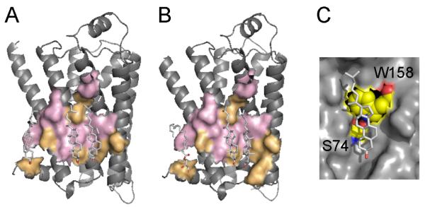
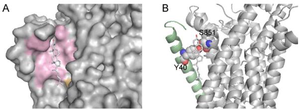
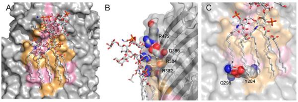
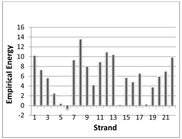
Membrane proteins function in the diverse environment of the lipid bilayer. Experimental evidence suggests that some lipid molecules bind tightly to specific sites on the membrane protein surface. These lipid molecules often act as co-factors and play important functional roles. In this study, we have assessed the evolutionary selection pressure experienced at lipid-binding sites in a set of α-helical and β-barrel membrane proteins using posterior probability analysis of the ratio of synonymous vs. nonsynonymous substitutions (ω-ratio). We have also carried out a geometric analysis of the membrane protein structures to identify residues in close contact with co-crystallized lipids. We found that residues forming cholesterol-binding sites in both β(2)-adrenergic receptor and Na(+)-K(+)-ATPase exhibit strong conservation, which can be characterized by an expanded cholesterol consensus motif for GPCRs. Our results suggest the functional importance of aromatic stacking interactions and interhelical hydrogen bonds in facilitating protein-cholesterol interactions, which is now reflected in the expanded motif. We also find that residues forming the cardiolipin-binding site in formate dehydrogenase-N γ-subunit and the phosphatidylglycerol binding site in KcsA are under strong purifying selection pressure. Although the lipopolysaccharide (LPS)-binding site in ferric hydroxamate uptake receptor (FhuA) is only weakly conserved, we show using a statistical mechanical model that LPS binds to the least stable FhuA β-strand and protects it from the bulk lipid. Our results suggest that specific lipid binding may be a general mechanism employed by β-barrel membrane proteins to stabilize weakly stable regions. Overall, we find that the residues forming specific lipid binding sites on the surfaces of membrane proteins often experience strong purifying selection pressure.


Figures

Fig. 1
Fig. 1
Cholesterol-binding sites in β2-adrenergic receptor shown in (A) 2RH1 and (B) 3D4S structures. Conserved residues are shown in pink, non-conserved residues are in orange. Cholesterol molecules bind in a shallow groove formed by the segments of helices I, II, III, and IV in similar, but not identical locations in both structures. The cholesterol-binding sites in both structures share five residues under strong purifying selection (shown in pink): I55, S74, C77, L80, L84, and W158. (C) The interhelical hydrogen bond between S74 from helix II and W158 from helix IV helps to maintain the optimal conformation of W158 side chain, which interacts with the sterol ring of cholesterol through the stacking interaction.
Fig. 2
Fig. 2
Cholesterol-binding site in Na+-K+-ATPase. (A) Protein surfaces formed by the residues under strong purifying selection pressure from α- and β-subunits are shown in pink. Non-conserved T788 from α-subunit is shown in orange. (B) Intersubunit hydrogen bond between Y40 (β-subunit, the helix is shown in pale green) and S851 (α-subunit) in the cholesterol-binding site, which promotes a stacking interaction with the bound cholesterol.
Fig. 3
Fig. 3
Lipopolysaccharide binding site in ferric hydroxamate uptake receptor. (A) The LPS-binding surface, where 11 residues are under strong purifying selection pressure, are shown in pink. The conserved residues cluster into two regions: one is a cluster of residues interacting with the polar saccharide head-group, and another cluster of residues interacting with the acyl chains in the hydrophobic core of the outer membrane. (B) Salt bridge interactions between conserved charged arginines and the LPS head-group. (C) Interstrand hydrogen bond between conserved Q298 and Y284, which may help to define the surface interacting with the acyl chain of phospholipid and provide additional interstrand stability.
Fig. 4
Fig. 4
Iindividual empirical β-strand energy calculations of the FhuA receptor. Strands 7 - 9 form the most unstable region in the protein, and β-strand 8, which runs through the middle of the LPS-binding site, has the highest energy.
Fig. 5
Fig. 5
Cardiolipin at the protein-protein interface in formate dehydrogenase N (Fdh-N). (A) Fragments of two Fdh-N heterotrimers are shown, where cardiolipin binds to the β and γ-subunits of one heterotrimer (shown in green and blue, respectively), and the γ–subunit from the adjacent heterotrimer (magenta). The bound heme molecules are also shown. Only one β-subunit is shown for clarity (colored in green). Here, cardiolipin (CL) acyl chain A fills in a tunnel leading to the heme-binding site, acyl chain B interacts with the neighboring γ-subunit, and acyl chain C interacts with β-subunit. (B) There are two CL-binding sites on the γ-subunit (shown in cyan and pink). In the first binding site, the acyl chain fills in the tunnel leading to the heme, and the majority of the tunnel residues are conserved (shown in pink). The second binding site is at the protein-protein interface with a neighboring γ-subunit. This site is the most conserved with the lowest ω-ratio, where all but one residue are under strong purifying selection. (C) CL-binding site on the β-subunit, which is shown as a green surface. The periplasmic loop residues N15, S16, and I16 form hydrogen bonds with the CL head-group, although they are not conserved. A263 and I266 that form a groove on the surface of TM helix are also not conserved.
Fig. 6
Fig. 6
Cardiolipin (CL) binding sites in ADP/ATP carrier. The conserved and non-conserved surfaces are shown in gray and red, respectively. (A, D) A front view (A) of the binding surface for cardiolipin CDL800 (the numbering is from 2C3E structure), mean ω = 0.033, and a view of the conserved tunnel (D) on the surface interacting with the CL head-group. The backbone carboxyl from G72 interacts with the phosphate group of the CL. (B, E) A front view (B) of the binding surface for cardiolipin CDL801, mean ω = 0.049, and a view of the conserved tunnel (E) interacting with the CL head-group. The backbone carboxyl from G272 interacts with the phosphate group. (C, F) A front view (C) of the binding surface for cardiolipin CDL802, mean ω = 0.022, and a view of the conserved tunnel (F) interacting with the CL head-group. The backbone carboxyl from G175 interacts with the phosphate group. (G) A sequence alignment of the five consecutive residues forming the bottom of the conserved depressions interacting with the cardiolipin head-groups. Each sequence contains a conserved aromatic residue (W70, Y173, and F270), and a conserved glycine (G72, G175, and G272). There are polar residues between the aromatics residues and the glycines: R71, Q174, and K271.
Fig. 7
Fig. 7
A phosphatidylglycerol (PG) binding site in KcsA potassium channel. The PG binding site is highly conserved with 9 out of 13 lipid-binding residues under strong purifying selection (shown in pink). R64, which was proposed [4] to interact with the negatively charged PG phosphate, is colored in blue.

References

    1. Singer SJ, Nicolson GL. The fluid mosaic model of the structure of cell membranes. Science. 1972;175:720–731. - PubMed
    1. Engelman DM. Membranes are more mosaic than fluid. Nature. 2005;438:578–580. - PubMed
    1. Dowhan W, Bogdanov M. Lipid-dependent membrane protein topogenesis. J. Ann. Rev. Biochem. 2009;78:515–540. - PMC - PubMed
    1. Valiyaveetil FI, Zhou YF, Mackinnon R. Lipids in the structure, folding, and function of the KcsA K+ channel. Biochemistry. 2002;41:10771–10777. - PubMed
    1. Cornelius F, Turner N, Christensen HRZ. Modulation of Na,K-ATPase by phospholipids and cholesterol. II. steady-state and presteady-state kinetics. Biochemistry. 2003;42:8541–8549. - PubMed

Publication types

MeSH terms

LinkOut - more resources