

Characterization of biomodified dentin matrices for potential preventive and reparative therapies
- PMID: 21167964
- PMCID: PMC3050116
- DOI: 10.1016/j.actbio.2010.12.013
Characterization of biomodified dentin matrices for potential preventive and reparative therapies
Abstract
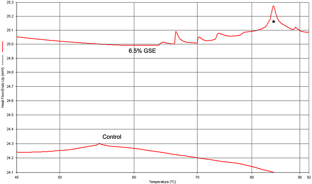
Biomodification of existing hard tissue structures, specifically tooth dentin, is an innovative approach proposed to improve the biomechanical and biochemical properties of tissue for potential preventive or reparative therapies. The objectives of the study were to systematically characterize dentin matrices biomodified by proanthocyanidin-rich grape seed extract (GSE) and glutaraldehyde (GD). Changes to the biochemistry and biomechanical properties were assessed by several assays to investigate the degree of interaction, biodegradation rates, proteoglycan interaction, and effect of collagen fibril orientation and environmental conditions on the tensile properties. The highest degree of agent-dentin interaction was observed with GSE, which exhibited the highest denaturation temperature, regardless of the agent concentration. Biodegradation rates decreased remarkably following biomodification of dentin matrices after 24h collagenase digestion. A significant decrease in the proteoglycan content of GSE-treated samples was observed using a micro-assay for glycosaminoglycans and histological electron microscopy, while no changes were observed for GD and the control. The tensile strength properties of GD-biomodified dentin matrices were affected by dentin tubule orientation, most likely due to the orientation of the collagen fibrils. Higher and/or increased stability of the tensile properties of GD- and GSE-treated samples were observed following exposure to collagenase and 8 months water storage. Biomodification of dentin matrices using chemical agents not only affects the collagen biochemistry, but also involves interaction with proteoglycans. Tissue biomodifiers interact differently with dentin matrices and may provide the tissue with enhanced preventive and restorative/reparative abilities.
Copyright © 2010 Acta Materialia Inc. Published by Elsevier Ltd. All rights reserved.
Figures


References
-
- Butler WT. Dentin matrix proteins and dentinogenesis. Connect Tissue Res. 1995;33:59–65. - PubMed
-
- Cheng H, Caterson B, Neame PJ, Lester GE, Yamauchi M. Differential distribution of lumican and fibromodulin in tooth cementum. Connect Tissue Res. 1996;34:87–96. - PubMed
-
- Beniash E, Traub W, Veis A, Weiner S. A transmission electron microscope study using vitrified ice sections of predentin: structural changes in the dentin collagenous matrix prior to mineralization. J Struct Biol. 2000;132:212–225. - PubMed
-
- Veis S, Sabsay B, Wu CB. Surface Reactive Peptides and Polymers. Washington, DC: 1991.
Publication types
MeSH terms
Substances
Grants and funding
LinkOut - more resources
Full Text Sources
Medical
