. 2011 Jan 4;108(1):355-60.
doi: 10.1073/pnas.1016785108. Epub 2010 Dec 15.

Multinodal regulation of the arcuate/paraventricular nucleus circuit by leptin

Affiliations

Multinodal regulation of the arcuate/paraventricular nucleus circuit by leptin

Masoud Ghamari-Langroudi et al. Proc Natl Acad Sci U S A. .

Abstract

Melanocortin-4 receptor (MC4R) is critical for energy homeostasis, and the paraventricular nucleus of the hypothalamus (PVN) is a key site of MC4R action. Most studies suggest that leptin regulates PVN neurons indirectly, by binding to receptors in the arcuate nucleus or ventromedial hypothalamus and regulating release of products like α-melanocyte-stimulating hormone (α-MSH), neuropeptide Y (NPY), glutamate, and GABA from first-order neurons onto the MC4R PVN cells. Here, we investigate mechanisms underlying regulation of activity of these neurons under various metabolic states by using hypothalamic slices from a transgenic MC4R-GFP mouse to record directly from MC4R neurons. First, we show that in vivo leptin levels regulate the tonic firing rate of second-order MC4R PVN neurons, with fasting increasing firing frequency in a leptin-dependent manner. We also show that, although leptin inhibits these neurons directly at the postsynaptic membrane, α-MSH and NPY potently stimulate and inhibit the cells, respectively. Thus, in contrast with the conventional model of leptin action, the primary control of MC4R PVN neurons is unlikely to be mediated by leptin action on arcuate NPY/agouti-related protein and proopiomelanocortin neurons. We also show that the activity of MC4R PVN neurons is controlled by the constitutive activity of the MC4R and that expression of the receptor mRNA and α-MSH sensitivity are both stimulated by leptin. Thus, leptin acts multinodally on arcuate nucleus/PVN circuits to regulate energy homeostasis, with prominent mechanisms involving direct control of both membrane conductances and gene expression in the MC4R PVN neuron.


Conflict of interest statement

The authors declare no conflict of interest.

Figures

Fig. 1.
Fig. 1.
Leptin tonically inhibits firing of MC4R PVN neurons. (A) Average ± SEM of action potential, firing frequency, and frequency distribution (i) of MC4R PVN neurons obtained from mice subjected to 16 h of fasting (n = 134) or fed ad libitum (n = 116, *P < 0.005). (B) Average ± SEM of firing frequency and frequency distribution (ii) of these neurons from 16-h fasted mice that were injected i.p. 3 h before decapitation with 3 μg/g leptin (n = 100) or saline (n = 99, *P < 0.005). (C) Average ± SEM of frequency of firing of MC4R PVN neurons obtained from ob/ob mice that were subjected to 16 h of fasting (n = 105), fed ad libitum (n = 132, P > 0.5), or injected i.p. with leptin 3 h before decapitation after 16 h of fasting (n = 100, *P < 0.0001).
Fig. 2.
Fig. 2.
α-MSH augments and AgRP and NPY decrease firing activity of MC4R PVN neurons. (A) Bath application of 250 nM α-MSH augments firing frequency of MC4R PVN neurons recorded using the loose-patch technique. (B) Average ± SEM of effect of 250 nM concentration of this peptide obtained from 13 MC4R PVN neurons (*P < 0.001). (C) Bath application of 100 nM AgRP significantly inhibits firing frequency of PVN neurons obtained by loose-patch recordings. (D) Average ± SEM of effect of 100 nM AgRP from seven PVN neurons (*P < 0.01). (E) A whole-cell recording from a spontaneously firing PVN neuron indicates that application of 100 nM NPY generates a significant inhibition of firing activity associated with hyperpolarization of membrane potential. (F and G) Average ± SEM of effect of 100 nM NPY on firing frequency (*P < 0.05) and membrane potentials (*P < 0.001) of eight MC4R PVN neurons.
Fig. 3.
Fig. 3.
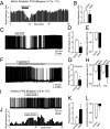
In vitro effects of leptin on firing activity of MC4R neurons in the mid- to posterior and anterior PVN. (A) Frequency histogram of effects of bath application of 50 nM leptin on firing activity of a MC4R PVN neuron recorded by loose-patch technique. (B) Average ± SEM of this effect obtained from 14 neurons recorded from mid- to posterior PVN (n = 14, *P < 0.0001). (C) A whole-cell recording indicates effects of 50 nM leptin on firing activity and membrane potentials of a spontaneously firing MC4R PVN neuron. (D and E) Average ± SEM of effects of bath application of 35–50 nM leptin on firing frequency (*P < 0.0001) and membrane potentials (*P < 0.0005) of 17 mid- to posterior MC4R PVN neurons. (F) A whole-cell recording indicates effects of 50 nM leptin on firing activity and membrane potential of a MC4R PVN neuron pretreated with 200 μM PTX and 1 mM KYN. (G and H) Average ± SEM of effects of 35–50 nM bath applied leptin on firing frequency (*P < 0.001) and membrane potential (*P < 0.05) of nine mid- to posterior MC4R PVN neurons pretreated with 200 μM PTX and 1 mM KYN. (I–L) Leptin activates action-potential firing activity of MC4R neurons in anterior PVN. (I) A whole-cell recording from a spontaneously firing MC4R neuron indicates that bath application of 35 nM leptin induces an increase in firing activity associated with depolarization of membrane potential. (J) The frequency histogram of this effect. (K and L) The bar graphs indicate average ± SEM of effects of 35–50 nM leptin on action-potential firing frequency (K) and membrane potential (L) of seven neurons tested (in both K and L, *P < 0.005).
Fig. 4.
Fig. 4.
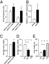
Fasting reduces responsiveness of MC4R PVN neurons to bath application of α-MSH in vitro. (A) Effect of bath applications of 250 nM α-MSH on firing frequency of MC4R PVN neurons obtained from 16-h fasted mice injected 3 h before decapitation with either 3 mg/kg leptin or saline (one-way ANOVA, *P = 0.0002). (B) Average ± SEM of magnitude of α-MSH–induced response indicates that this response is greater in neurons from mice treated with leptin (∼5.6-fold increase, n = 17) than those treated with saline (∼1.5-fold increase, n = 15, *P < 0.0005, one-way ANOVA). (C) Hypothalamic expression of MC4R gene normalized to β-actin gene obtained from fasted mice (20 h) that were injected i.p. 5 h before decapitation with either saline (n = 20) or leptin (n = 19, *P < 0.01). Data were obtained using quantitative real-time PCR. (D) Hypothalamic expression of MC4R normalized to β-actin gene obtained from MC4R−/− (KO, n = 5), MC4R−/+ (HET, n = 5), and MC4R+/+ (WT, n = 12) adult male mice fed with normal chow. Asterisk indicates all groups are significantly different (P < 0.0001, one-way ANOVA). Data were obtained using quantitative real-time PCR. (E) Average ± SEM of body weight of corresponding groups of mice as in D (*P < 0.0001, one-way ANOVA).

References

    1. Huszar D, et al. Targeted disruption of the melanocortin-4 receptor results in obesity in mice. Cell. 1997;88:131–141. - PubMed
    1. Fan W, Boston BA, Kesterson RA, Hruby VJ, Cone RD. Role of melanocortinergic neurons in feeding and the agouti obesity syndrome. Nature. 1997;385:165–168. - PubMed
    1. Cone RD. Anatomy and regulation of the central melanocortin system. Nat Neurosci. 2005;8:571–578. - PubMed
    1. Zhang Y, et al. Positional cloning of the mouse obese gene and its human homologue. Nature. 1994;372:425–432. - PubMed
    1. Fekete C, et al. α-Melanocyte-stimulating hormone is contained in nerve terminals innervating thyrotropin-releasing hormone-synthesizing neurons in the hypothalamic paraventricular nucleus and prevents fasting-induced suppression of prothyrotropin-releasing hormone gene expression. J Neurosci. 2000;20:1550–1558. - PMC - PubMed

Publication types

MeSH terms

LinkOut - more resources