. 2011 Feb 25;286(8):6006-16.
doi: 10.1074/jbc.M110.198796. Epub 2010 Dec 17.

Preferential repair of oxidized base damage in the transcribed genes of mammalian cells

Affiliations

Preferential repair of oxidized base damage in the transcribed genes of mammalian cells

Dibyendu Banerjee et al. J Biol Chem. .

Abstract

Preferential repair of bulky DNA adducts from the transcribed genes via nucleotide excision repair is well characterized in mammalian cells. However, definitive evidence is lacking for similar repair of oxidized bases, the major endogenous DNA lesions. Here we show that the oxidized base-specific human DNA glycosylase NEIL2 associates with RNA polymerase II and the transcriptional regulator heterogeneous nuclear ribonucleoprotein-U (hnRNP-U), both in vitro and in cells. NEIL2 immunocomplexes from cell extracts preferentially repaired the mutagenic cytosine oxidation product 5-hydroxyuracil in the transcribed strand. In a reconstituted system, we also observed NEIL2-initiated transcription-dependent base excision repair of 5-hydroxyuracil in the transcribed strand, with hnRNP-U playing a critical role. Chromatin immunoprecipitation/reimmunoprecipitation studies showed association of NEIL2, RNA polymerase II, and hnRNP-U on transcribed but not on transcriptionally silent genes. Furthermore, NEIL2-depleted cells accumulated more DNA damage in active than in silent genes. These results strongly support the preferential role of NEIL2 in repairing oxidized bases in the transcribed genes of mammalian cells.


Figures

FIGURE 1.
FIGURE 1.
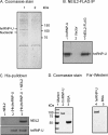
Co-IP analysis was performed by adding the relevant Ab to the DNase/RNase- or benzonase-treated extract as described earlier (26). A, extracts from AGS cells stably expressing NEIL2-FLAG (lane 1) or empty FLAG vector (lane 2) were immunoprecipitated with anti-FLAG Ab and tested for the presence of RNAP II by Western analysis with the N-20 Ab (Santa Cruz Biotechnology, Inc.). B, extracts from AGS cells stably expressing NEIL2-FLAG (lane 1) were immunoprecipitated with anti-RNAP II Ab (N-20; lane 3) or control IgG (lane 2) and tested for the presence of NEIL2-FLAG using anti-FLAG Ab. C, endogenous NEIL2 from the extract of normal AGS cells (lane 1) was immunoprecipitated using anti-NEIL2 Ab (lane 3) or IgG (lane 2) and tested for the presence of RNAP II using N20 Ab. D, the RNAP II (lane 2) or control IgG (lane 3) immunocomplex from the extract of the same cells (lane 1) was tested for the presence of endogenous NEIL2 using anti-NEIL2 Ab. Lane 4, purified NEIL2 as marker.
FIGURE 2.
FIGURE 2.
A, identification of NEIL2-associated proteins. Proteins bound to NEIL2-FLAG (lane 1) or empty vector (lane 2) were analyzed by SDS-PAGE and Coomassie blue staining. The indicated protein bands were identified by MS analysis. B, Western analysis to identify hnRNP-U in the NEIL2-FLAG immunoprecipitate (lane 3). Lane 2, vector immunoprecipitate as control; lane 1, purified hnRNP-U as a size marker. C, Ni2+-NTA pull-down of NEIL2 using purified His-tagged hnRNP-U. Ni2+-NTA beads (15 μl; Sigma) were washed thoroughly with buffer B (50 mm Tris-Cl, pH 7.5, 0.1% Triton X-100, and 1 mm DTT) containing 150 mm NaCl and incubated with His-hnRNP-U (90 pmol; lanes 2 and 3) in the wash buffer (200 μl) for 1 h. The beads were then washed extensively to remove all unbound proteins. hnRNP-U-bound beads (lane 3) or the beads alone (lane 1) were then incubated with untagged NEIL2 (45 pmol) for 45 min and washed to remove unbound proteins. Finally, the beads were washed with 500 mm NaCl containing buffer B and boiled with SDS loading dye, and the supernatant was analyzed by SDS-PAGE to identify the bound proteins by immunoblotting using anti-NEIL2 (top) or anti-hnRNP-U Ab (bottom). D, far Western analysis of hnRNP-U with NEIL2. Recombinant proteins were separated by SDS-PAGE in two separate gels, one of which was Coomassie-stained (lanes 1–3), whereas proteins in the other gel were transferred to a nitrocellulose membrane for interaction studies with NEIL2 (lanes 4 and 5) followed by immunostaining with anti-NEIL2 Ab.
FIGURE 3.
FIGURE 3.
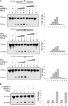
Stimulation of NEIL2 activity by hnRNP-U. A, bubble (5-OHU·B11; top), duplex (5-OHU·G; middle), or single-stranded (5-OHU; bottom) oligonucleotide (2 pmol) was incubated at 37 °C for 10 min with hnRNP-U (0.2 pmol; lane 2), NEIL2 (0.2 pmol; lane 3), or RPA alone (0.2 pmol; lane 10) or with NEIL2 (0.2 pmol) with increasing amounts of hnRNP-U (0.05, 0.1, and 0.2 pmol; lanes 4–6) or RPA (0.05, 0.1, and 0.2 pmol; lanes 7–9). S, substrate; P, product. The histograms show quantitation of the cleaved products, setting the activity of NEIL2 alone (lane 3) to 1. Values for these histograms and those in all other figures represent the mean from at least three independent experiments. B, specificity of the stimulatory activity of hnRNP-U. Two pmol of substrate DNA (5-OHU·B11) was incubated at 37 °C for 10 min with NEIL2 (0.2 pmol) in the absence (lane 4) or the presence (lane 5) of anti-hnRNP-U Ab or anti-hnRNP-U Ab plus excess hnRNP-U protein (++, lane 6). Lane 1, no protein; lanes 2 and 3, NEIL2 alone and NEIL2 plus anti-hnRNP-U Ab, respectively. The histograms show quantitation of the cleaved product relative to NEIL2 alone (lane 2). Error bars, S.E.
FIGURE 4.
FIGURE 4.
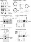
TC-BER using NEIL2-FLAG immunocomplex. A, partial characterization of the NEIL2-FLAG immunocomplex by Western blot analysis. Extracts from AGS cells stably expressing NEIL2-FLAG (lane 3) or vector alone (lane 4) were immunoprecipitated with anti-FLAG Ab and tested for the presence of NEIL2-associated proteins with Abs to the proteins shown on the right. B, scheme for analysis of transcription-coupled BER using plasmid DNA containing a single 5-OHU in the transcribed or non-transcribed strand (pUC-TS-5-OHU or -NTS-5-OHU, respectively). The plasmids contain an adenoviral late promoter (gray box); the lesion (*) was introduced 167 bp downstream of the transcription start site (shown with an arrowhead) (30). C, repair of pUC-TS-5-OHU (lanes 2 and 3 and lanes 6 and 7) or pUC-NTS-5-OHU (lanes 4 and 5) plasmid DNAs using NEIL2-FLAG (lanes 2–5) or FLAG-vector immunoprecipitate (lanes 6 and 7), with (+) or without (−) 250 μm NTPs. Lane 1, 5′-end-labeled EcoRI/HindIII fragment from pUC-TS plasmid DNA as a size marker. Other details are provided under “Experimental Procedures.” The repaired plasmid was cleaved with EcoRI/HindIII and analyzed in a 6% TBE-urea gel. Complete repair is indicated by radioactive dTMP incorporation in the plasmid fragment spanning the 5-OHU lesion site. The histograms show quantitation of the repaired product relative to that in the TS without NTPs (lane 3). D, effect of α-amanitin on NEIL2-initiated BER. The repair assays were carried out using NEIL2-FLAG immunoprecipitate in the presence (lane 2) or absence (lanes 1 and 3) of α-amanitin and in the absence (lane 1) or presence (lanes 2 and 3) of NTPs, respectively. Quantitation of the repaired product is shown in histograms at the bottom; repair activity without α-amanitin (lane 3) and with NTPs was set to 1. E, analysis of transcript. The NEIL2-FLAG immunocomplex was incubated for 1 h at 30 °C with 200 ng of pUC-TS-5-OHU (TS, lane 3) or pUC-NTS-5-OHU (NTS, lane 4) plasmid DNA (30) in a 50-μl reaction buffer (25 mm HEPES-KOH, pH 7.9, 50 mm KCl, 5 mm MgCl2, 0.5 mm DTT) containing 20 units of RNase inhibitor and a 250 μm concentration each of GTP, CTP, and ATP; 10 μm UTP; and 5 μCi of [α-32P]UTP. The reaction mixture was phenol/chloroform-extracted, ethanol-precipitated, and analyzed by denaturing gel electrophoresis. Lanes 1 and 2, marker DNAs 5′-labeled with [γ-32P]ATP. F, the specificity of hnRNP-U in NEIL2-initiated BER was tested using the NEIL2-FLAG immunocomplex either in the absence (lane 3) or presence (lanes 1 and 2) of anti-hnRNP-U Ab. The effect of the anti-hnRNP-U Ab could be partially reversed by the addition of purified hnRNP-U protein (0.2 pmol; lane 1). The histograms show quantitation of the repaired product relative to the repair activity of NEIL2-FLAG immunoprecipitate (lane 3). Lane 4, marker. Error bars, S.E.
FIGURE 5.
FIGURE 5.
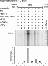
In vitro reconstitution of TC-BER. For radioactive incorporation into pUC-TS-5-OHU (with [α-32P]dTMP (lanes 1–7) or [α-32P]dAMP (lane 8)), 10 pmol of substrate was incubated with 0.5 pmol of NEIL2 (lanes 1 and 3–8), 25 fmol of PNK, 0.25 pmol each of Pol β and Lig IIIα, and an RNAP II fraction containing TBP, TFIIB, and E/F/H (Protein One, except in lane 5 without the transcription factors) in the presence (+) or absence (−) of NTPs, as described under “Experimental Procedures.” Lane 1, undamaged DNA; lane 9, end-labeled ϕX174 HinfI DNA marker (Promega). Complete repair is indicated by the [32P]dTMP incorporation. The repaired plasmid was linearized with EcoRI/HindIII and analyzed in a 6% TBE-urea gel; the gel was dried and the reaction products were analyzed and quantitated with a PhosphorImager. Bottom, histograms indicating the -fold increase in repair activity; activity without NTPs (lane 3) was set to 1. Error bars, S.E.
FIGURE 6.
FIGURE 6.
ChIP/re-ChIP assay. After sonication and immunoprecipitation of cross-linked chromatin with the first Ab (1st IP), the bound fraction was eluted, divided into two aliquots, and subjected to a second round of immunoprecipitation (2nd IP) with IgG (as control) or the specific Ab. A, ChIP/re-ChIP assay involving NEIL2-RNAPII or NEIL2-hnRNP-U. PCR amplifications were performed using the primers flanking the coding regions of actively transcribed or repressed genes in AGS and SK-N-BE2-(C) cells. Note that lanes 1 and 3 and lanes 6 and 8 each show corresponding assay pairs in the reciprocal order. B, ChIP/re-ChIP assay involving NEIL2 and APE1. Cross-linked chromatin was immunoprecipitated from NEIL2-FLAG-expressing AGS cell extract with anti-FLAG (lane 2) or anti-APE1 (lane 3) Ab, and the NEIL2-bound fraction was then subjected to a second round of immunoprecipitation with IgG (lane 4) or anti-APE1 (lane 5) Ab. PCR amplifications were performed using primers flanking the coding regions of β-actin, transcribed in all cell types. Other details are provided under “Experimental Procedures.”
FIGURE 7.
FIGURE 7.
Quantitation of DNA damage in NEIL2-depleted cells. A, Western analysis of NEIL2 in stably transfected control and NEIL2 shRNA-expressing cells. Top, RNAP II as control. B, the histogram depicts the relative increase in DNA damage level calculated from the increase in Ct values of 2.65 ± 0.42 and 2.27 ± 0.68 for the transcribed (β-actin and MYOD, respectively) and 1.08 ± 0.03 and 1.45 ± 0.11 for the nontranscribed genes (MASH and NEUROD, respectively) in NEIL2-depleted versus control cells. Error bars, S.E.

References

    1. Breimer L. H. (1990) Mol. Carcinog. 3, 188–197 - PubMed
    1. Lavrovsky Y., Chatterjee B., Clark R. A., Roy A. K. (2000) Exp. Gerontol. 35, 521–532 - PubMed
    1. Krokan H. E., Nilsen H., Skorpen F., Otterlei M., Slupphaug G. (2000) FEBS Lett. 476, 73–77 - PubMed
    1. Friedberg E. C., Walker G. C., Siede W., Wood R. D., Schultz R. A., Ellenberger T. (2006) DNA Repair and Mutagenesis, American Society for Microbiology Press, Washington, D. C.
    1. Ikeda S., Biswas T., Roy R., Izumi T., Boldogh I., Kurosky A., Sarker A. H., Seki S., Mitra S. (1998) J. Biol. Chem. 273, 21585–21593 - PubMed

Publication types

MeSH terms

LinkOut - more resources