. 2011 Feb 15;22(4):478-92.
doi: 10.1091/mbc.E10-03-0233. Epub 2010 Dec 17.

Huntingtin coordinates the dynein-mediated dynamic positioning of endosomes and lysosomes

Affiliations

Huntingtin coordinates the dynein-mediated dynamic positioning of endosomes and lysosomes

Juliane P Caviston et al. Mol Biol Cell. .

Abstract

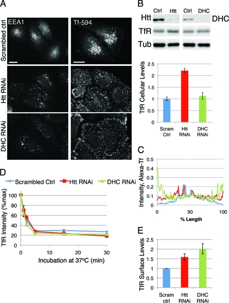
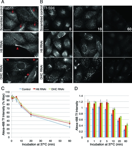
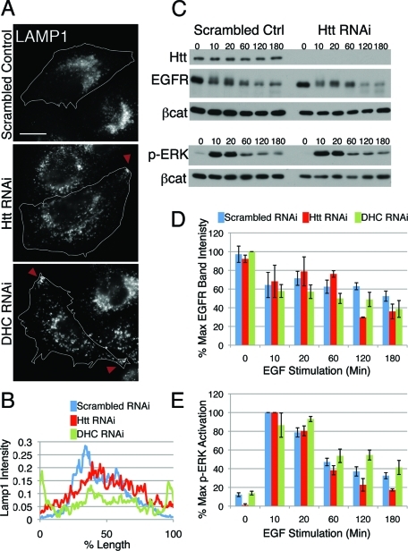
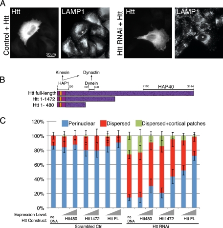
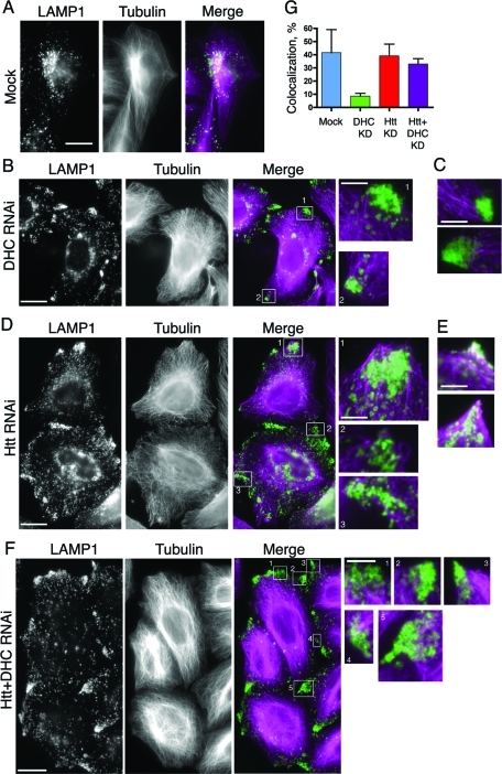
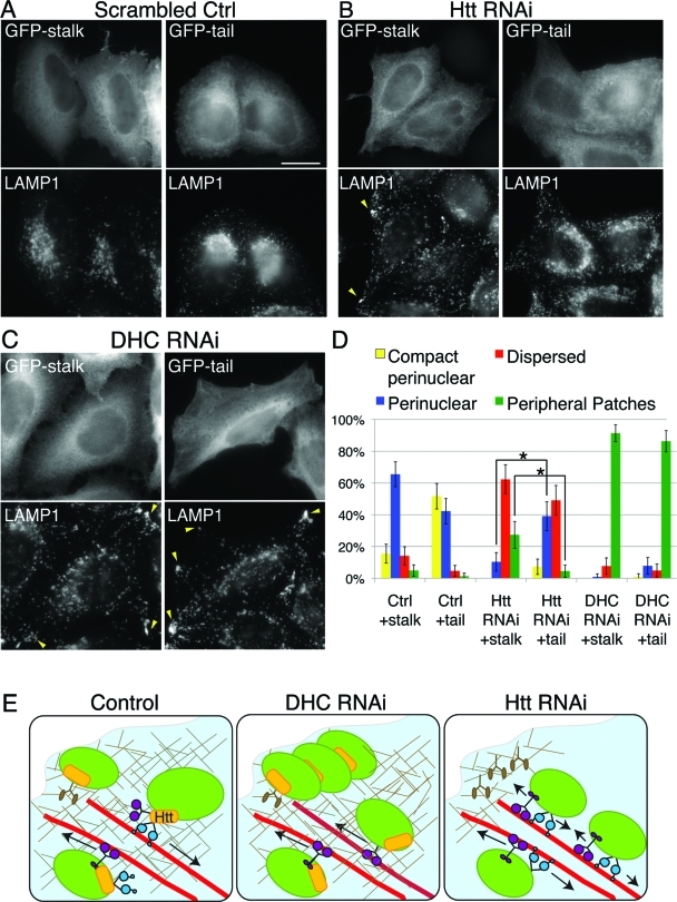
Huntingtin (Htt) is a membrane-associated scaffolding protein that interacts with microtubule motors as well as actin-associated adaptor molecules. We examined a role for Htt in the dynein-mediated intracellular trafficking of endosomes and lysosomes. In HeLa cells depleted of either Htt or dynein, early, recycling, and late endosomes (LE)/lysosomes all become dispersed. Despite altered organelle localization, kinetic assays indicate only minor defects in intracellular trafficking. Expression of full-length Htt is required to restore organelle localization in Htt-depleted cells, supporting a role for Htt as a scaffold that promotes functional interactions along its length. In dynein-depleted cells, LE/lysosomes accumulate in tight patches near the cortex, apparently enmeshed by cortactin-positive actin filaments; Latrunculin B-treatment disperses these patches. Peripheral LE/lysosomes in dynein-depleted cells no longer colocalize with microtubules. Htt may be required for this off-loading, as the loss of microtubule association is not seen in Htt-depleted cells or in cells depleted of both dynein and Htt. Inhibition of kinesin-1 relocalizes peripheral LE/lysosomes induced by Htt depletion but not by dynein depletion, consistent with their detachment from microtubules upon dynein knockdown. Together, these data support a model of Htt as a facilitator of dynein-mediated trafficking that may regulate the cytoskeletal association of dynamic organelles.


Figures

FIGURE 1:
FIGURE 1:
Htt is required for perinuclear early endosome positioning but not for transferrin uptake from the plasma membrane. (A) HeLa cells transfected with scrambled control oligo (top), Htt RNAi (middle), or DHC RNAi (bottom) were stained with antibodies against EEA1 (left column) or given a quick pulse-chase with 594-Alexa-Tf (right column) to label early endosomes. Scale bar = 20 μm. (B) Top: Western blot demonstrating decreased levels of Htt (top left panel) and DHC (top right panel) in RNAi cells. TfR is detected in the middle panel and tubulin is shown as a loading control. Bottom: Due to the near-saturation levels of tubulin immunoreactivity, TfR levels were normalized using β-catenin (not shown) as a loading control. (C) Linescans of control (n = 12), Htt RNAi (n = 11), and DHC RNAi (n = 12) cells pulsed with Tf-594 (see A), with average fluorescence intensity along the length of the cell. (D) Control, Htt RNAi, and DHC RNAi cells were incubated with antibody to TfR at 4°C, then shifted to 37°C to allow internalization for 0, 1, 2, 5, 15, and 30 min; incubated with fluorescent secondary antibody; then fixed and processed for flow cytometry. Average fluorescent intensity of TfR is plotted as percent of maximum (n = 4). (E) TfR present on the surface is measured as fluorescence intensity at the zero time point for scrambled control, Htt RNAi, and DHC RNAi cells, normalized by the mean intensity for scrambled control cells. Error bars, ±SEM.
FIGURE 2:
FIGURE 2:
Recycling compartments in cells depleted of Htt or dynein lack perinuclear concentration, but kinetics of recycling are normal. (A) Scrambled control cells (top), Htt RNAi cells (middle), and DHC RNAi cells stained for Rab11. Concentrations of Rab11 at the perinucleus are denoted with arrows, and peripheral accumulations are denoted by arrowheads. (B) Cells were pulsed with Alexa594-Tf for 30 min to label recycling compartments, washed extensively, and then transferred to regular media and fixed at various time points. Shown are images of scrambled control (first row), Htt RNAi (second row), and DHC RNAi (third row) cells fixed at 0 (left), 10 (middle), and 60 (right) min. (C) Cells were treated as above but with Alexa 488-Tf, and cell intensities at 0, 1, 2, 5, 10, 20, and 60 min were measured by flow cytometry. Average fluorescence intensity of 488-Tf is plotted as percent of maximum. Htt RNAi, n = 3; DHC RNAi, n = 4; scrambled control, n = 7. (D) Alexa 488-Tf intensities at each time point of (C), normalized by the mean fluorescence intensity of scrambled control. All scale bars = 20 μm. Error bars, ±SEM.
FIGURE 3:
FIGURE 3:
LE/lysosomes are mislocalized in Htt RNAi cells; however, degradative function is normal. (A) LE/lysosomes in scrambled control (top), Htt RNAi (middle), and DHC RNAi cells (bottom) were visualized by staining cells for LAMP1. Peripheral accumulations are denoted with arrowheads. Scale bar = 20 μm. (B) Linescans of LAMP1 intensity in scrambled control (blue; n = 13), Htt RNAi (red; n = 15), and DHC RNAi cells (green; n = 16). (C) Western blot of scrambled control and Htt RNAi cells stimulated with EGF for various times. Cell lysates were probed for Htt, EGFR, and phospho-ERK1/2 with β-catenin as a loading control. The experiment was done in triplicate; a representative image is shown. (D, E) Densitometry of EGFR bands (D) and p-ERK1/2 bands (E) in scrambled control, Htt RNAi, and DHC RNAi cell lysates throughout the EGF stimulation, normalized for loading with β-catenin. EGF stimulation of DHC RNAi cells was performed three times in duplicate. Error bars, ±SEM.
FIGURE 4:
FIGURE 4:
Full-length Htt is required for perinuclear organelle positioning. (A) Scrambled control (left) or Htt RNAi cells (right) were transfected with full-length Htt and stained for Htt to identify transfected cells and LAMP1. Asterisks identify transfected cells in LAMP1 panels. Scale bar = 20 μm. (C) Schematic of Htt full-length, Htt aa1–1472, and Htt aa1–480. The polyglutamine repeat region begins at aa17 (orange), and there are 36 predicted HEAT repeats spanning the length of the peptide (dark purple speckled). HAP1, which associates with kinesin and dynactin, binds to the Htt N terminus, dynein binds aa601–698, and HAP40 binding site resides at the C terminus. (D) Scrambled control (left side) or Htt RNAi (right side) cells transfected with no DNA, Htt1–480, Htt1–1472, or Htt full-length (Htt FL) were scored for perinuclear (blue), dispersed (red), or dispersed with additional cortical accumulations of LE/lysosomes (green), visualized by staining for LAMP1. To illustrate the change in phenotypes at different Htt construct expression levels, the transfected cells were binned into low or medium expressors vs. high expressors; expression level is signified by the gray ramp; n ≥ 64 cells for each condition. Error bars, ±95% CI.
FIGURE 5:
FIGURE 5:
Htt may play a role in coordinating organelle association with microtubules. HeLa cells were mock transfected (A) or depleted of DHC (B) and stained for LE/lysosomes (LAMP1) and microtubules. The magnified boxed regions (boxes 1 and 2) show that the peripheral LE/lysosomes are most often found just beyond the tips of the microtubules at the cell periphery. (C) Additional merged images (as in B) featuring magnified regions of peripheral LE/lysosomes and microtubules. (D) HeLa cells depleted of Htt were stained as in (A). The magnified boxed regions (boxes 1–3) show that the peripheral LE/lysosomes are most often found enmeshed in regions dense with microtubules. (E) Additional merged images (as in D) featuring magnified regions of overlapping LE/lysosomes and microtubules at the cell edge. (F) HeLa cells depleted of Htt and dynein were stained as in (A). The magnified boxed regions (boxes 1–4) show that the peripheral LE/lysosomes are most often found enmeshed in regions dense with microtubules, similar to (D). Scale bar = 20 μm, except magnified images. The magnified images are 4× the size of original image; scale bar = 5 μm. (G) Colocalization of peripheral patches of LE/lysosomes and microtubules was quantified in mock (n = 4), DHC RNAi (n = 17), Htt RNAi (n = 4), and Htt and DHC double-knockdown cells (n = 16). Error bars, ±SEM.
FIGURE 6:
FIGURE 6:
Peripheral lysosomes are distributed in regions enriched for cortical actin in cells depleted for dynein. (A) Mock-treated control cells were stained with antibody to LAMP-1 to visualize LE/lysosomes and with phalloidin to visualize F-actin. Boxed regions 1–2 highlight very tiny patches of peripheral LE/lysosomes. (B) DHC RNAi cells were stained for LE/ lysosomes (LAMP1) and F-actin. Boxed regions (1 and 2) in the merged image show that lysosomal accumulations on the cell periphery (green) are present in regions enriched with cortical actin (magenta). (C) Htt RNAi cells were stained as in (B). Boxed regions (1 and 2) in the merged image show that lysosomal accumulations on the cell periphery (green) are in areas with less marked staining for F-actin (magenta). Scale bar = 20 μm, except magnified images. The magnified images are 4× the size of original image; scale bar = 5 μm.
FIGURE 7:
FIGURE 7:
A dynamic actin network surrounds LE/lysosomes on cell periphery in dynein-depleted cells, but not Htt-depleted cells. (A) Cells transfected with control RNAi oligos (top row) and cells depleted of Htt (second row), DHC (third row), or Htt and DHC double-knockdown cells were stained for LAMP1 (first column) or cortactin (middle column). Boxes 1–4 are magnified regions highlighting the cortactin meshwork surrounding the peripheral LE/lysosomes in DHC RNAi cells, which is absent in Htt RNAi or Htt and DHC double-knockdown cells. Scale bar = 20 μm, except magnified images. The magnified images are 4× the size of original image; scale bar = 5 μm. (B) The sizes of the peripheral LE/lysosomal patches were evaluated in mock (n = 4), Htt RNAi (n = 7), DHC RNAi (n = 37), or Htt and DHC double-knockdown cells (n = 37). Error bars, ±SEM.
FIGURE 8:
FIGURE 8:
Peripheral LE/lysosomal patches induced by dynein depletion are lost upon depolymerization of actin filaments with Latrunculin B. Cells were depleted of either dynein or Htt by siRNA, or were treated with scrambled control oligos, then were treated with either DMSO (left) or Latrunculin B. Prominent peripheral LAMP1-positive patches formed in dynein RNAi cells are not seen in drug-treated cells. Latrunculin-induced depolymerization of actin filaments was visualized with phalloidin staining. Scale bar = 20 μm.
FIGURE 9:
FIGURE 9:
LE/lysosomal dispersion in Htt knockdown cells is mitigated by disruption of kinesin activity in Htt-knockdown but not DHC-knockdown cells. (A–C) Scrambled control (A), Htt RNAi (B), or DHC RNAi (C) cells were transfected with GFP-KIF5C stalk (GFP-stalk, left panel) or GFP-KIF5C tail (GFP-tail, right panel). LE/lysosomes of transfected cells were visualized by staining for LAMP1. Note that, in (B), transfection of GFP-tail restores a perinuclear concentration of LE/lysosomes that is absent in cells transfected with the control plasmid, GFP-stalk. In (C), peripheral accumulations of LE/lysosomes (arrowheads) persist in DHC RNAi cells in GFP-tail–positive cells. Scale bar = 20 μm. (D) Percentage of total cells transfected with GFP-tail or GFP-stalk that exhibited compact perinuclear (yellow), perinuclear (blue), dispersed (red), or peripheral LE/lysosomes (green). Error bars, ±95% CI. *, p ≤ 0.0001. (p values generated using Fisher’s exact test for proportional data.) (E) Model of a role for Htt in coordinating organelle transport along the actin and microtubule cytoskeletons. In control cells, vesicular cargo, such as LE/lysosomes (green), are transported to the cell periphery along the microtubule (red) by kinesin-1 (purple) possibly via an adaptor complex involving HAP1 and Htt (yellow). There, they undergo a Htt-dependent change in cytoskeletal affinity, possibly via a myosin (brown) linker molecule such as optineurin. LE/lysosomes are able to return to the cell center by dynein-dependent microtubule transport (blue), facilitated by Htt. Under DHC RNAi conditions, LE/lysosomes can no longer undergo minus-end–directed microtubule transport and thus accumulate along the cell edge in the cortical actin network and are enmeshed in newly polymerized actin. In Htt RNAi cells, there is a decrease in new actin along the cell cortex, a defect in switching cytoskeletal tracks and inefficient dynein-mediated microtubule transport. As a result, LE/lysosomes do not concentrate at the perinuclear region and instead become more evenly distributed throughout the cytoplasm and accumulate at the microtubule plus-ends.

References

    1. Aridor M, Hannan LA. Traffic jam: a compendium of human diseases that affect intracellular transport processes. Traffic. 2000;1:836–851. - PubMed
    1. Aridor M, Hannan LA. Traffic jams II: an update of diseases of intracellular transport. Traffic. 2002;3:781–790. - PubMed
    1. Atwal RS, Xia J, Pinchev D, Taylor J, Epand RM, Truant R. Huntingtin has a membrane association signal that can modulate huntingtin aggregation, nuclear entry and toxicity. Hum Mol Genet. 2007;16:2600–2615. - PubMed
    1. Brown CL, Maier KC, Stauber T, Ginkel LM, Wordeman L, Vernos I, Schroer TA. Kinesin-2 is a motor for late endosomes and lysosomes. Traffic. 2005;6:1114–1124. - PubMed
    1. Burkhardt JK, Echeverri CJ, Nilsson T, Vallee RB¡. Overexpression of the dynamitin (p50) subunit of the dynactin complex disrupts dynein-dependent maintenance of membrane organelle distribution. J Cell Biol. 1997;139:469–484. - PMC - PubMed

Publication types

MeSH terms