. 2011 Jan;18(1):41-4.
doi: 10.1107/S0909049510036022. Epub 2010 Nov 5.

Biomolecular solution X-ray scattering at the National Synchrotron Light Source

Affiliations

Biomolecular solution X-ray scattering at the National Synchrotron Light Source

Marc Allaire et al. J Synchrotron Radiat. 2011 Jan.

Abstract

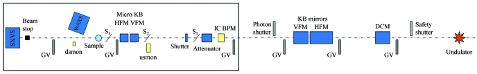
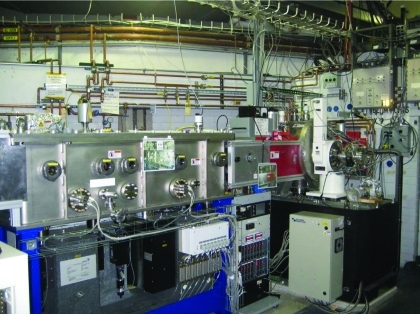
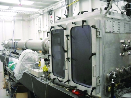
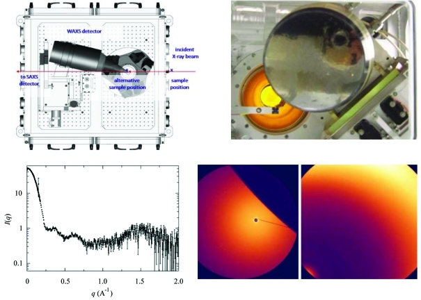
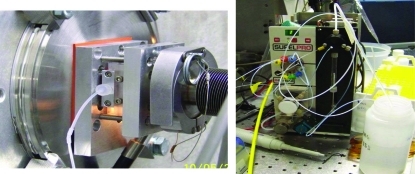
In recent years there has been a growing interest in the application of X-ray scattering techniques to biomolecules in solution. At NSLS, a new undulator-based beamline, X9, has been constructed to address the oversubscribed user demand for X-ray scattering. Beamline X9 has the capability to perform small/wide-angle X-ray scattering (SAXS/WAXS) all in one single instrument. This is accomplished by utilizing a vacuum sample/detector chamber that is an integral part of the SAXS scattering flight path. This vacuum chamber allows a WAXS detector to be positioned at a close distance from the sample, while not interfering with scattered X-rays at small angles from reaching the SAXS detector. A regular training program, the X9 workbench, has also been established to allow users to become familiar with beamline X9 for solution X-ray scattering.


Figures

Figure 1
Figure 1
Schematic of the principal components of the NSLS beamline X9 from the undulator source (at the right) to the endstation hutch (large black box). Abbreviations used: gate valve (GV); double-crystal monochromator (DCM); Kirkpatrick–Baez (KB); horizontal/vertical focusing mirrors (HFM/VFM); ion chamber beam-position monitor (IC BPM); slits (S1, S2, S3); upstream/downstream monitor (usmon/dsmon).
Figure 2
Figure 2
Photograph of the beamline optical components with the double-crystal monochromator (on the right) and the KB horizontal/vertical bimorph adaptive focusing mirrors (on the left).
Figure 3
Figure 3
The long vacuum-compatible chamber of NSLS beamline X9 that contains both the SAXS (at the far end) and WAXS detectors (designed by Scott Coburn).
Figure 4
Figure 4
The NSLS X9 beamline was especially designed to acquire both SAXS and WAXS X-ray scattering data from a single exposure. The top-left panel indicates the position of the WAXS and the SAXS detectors relative to the X-ray beam. The top-right panel is a view from the X-ray beam in the direction of the detectors. This view indicates the overlapping region, which can be seen from the SAXS images (bottom-right panel, left image) cover by the WAXS detector (bottom-right panel, right image). The SAXS and WAXS data overlap between ∼0.12 and 0.2 Å−1 and are background-subtracted and combined automatically using beamline software, as shown by the data collected from a 3.7 mg ml−1 lysozyme solution (bottom-left).
Figure 5
Figure 5
The solution sample cell at NSLS beamline X9 using a 1 mm glass capillary sealed across the vacuum path (left). The sample cell is connected through standard fittings to a Kloehn Versa 3 syringe pump (right).

References

    1. Jackques, D. A. & Trewhella, J. (2010). Protein Sci.19, 642–657. - PMC - PubMed
    1. Makowski, L. (2010). J. Struct. Funct. Genom.11, 9–19. - PMC - PubMed
    1. Mertens, H. D. T. & Svergun, D. I. (2010). J. Struct. Biol.172, 128–141. - PubMed
    1. Rambo, R. P. & Tainer, J. A. (2010). Curr. Opin. Struct. Biol.20, 128–137. - PMC - PubMed

Publication types