. 2011 Mar-Apr;17(3-4):256-65.
doi: 10.2119/molmed.2010.00097. Epub 2010 Dec 15.

Differential effects of HNF-1α mutations associated with familial young-onset diabetes on target gene regulation

Affiliations

Differential effects of HNF-1α mutations associated with familial young-onset diabetes on target gene regulation

Maria Galán et al. Mol Med. 2011 Mar-Apr.

Abstract

Hepatocyte nuclear factor 1-α (HNF-1α) is a homeodomain transcription factor expressed in a variety of tissues (including liver and pancreas) that regulates a wide range of genes. Heterozygous mutations in the gene encoding HNF-1α (HNF1A) cause familial young-onset diabetes, also known as maturity-onset diabetes of the young, type 3 (MODY3). The variability of the MODY3 clinical phenotype can be due to environmental and genetic factors as well as to the type and position of mutations. Thus, functional characterization of HNF1A mutations might provide insight into the molecular defects explaining the variability of the MODY3 phenotype. We have functionally characterized six HNF1A mutations identified in diabetic patients: two novel ones, p.Glu235Gly and c-57-64delCACGCGGT;c-55G>C; and four previously described, p.Val133Met, p.Thr196Ala, p.Arg271Trp and p.Pro379Arg. The effects of mutations on transcriptional activity have been measured by reporter assays on a subset of HNF-1α target promoters in Cos7 and Min6 cells. Target DNA binding affinities have been quantified by electrophoretic mobility shift assay using bacterially expressed glutathione-S-transferase (GST)-HNF-1α fusion proteins and nuclear extracts of transfected Cos7 cells. Our functional studies revealed that mutation c-57-64delCACGCGGT;c-55G>C reduces HNF1A promoter activity in Min6 cells and that missense mutations have variable effects. Mutation p.Arg271Trp impairs HNF-1α activity in all conditions tested, whereas mutations p.Val133Met, p.Glu235Gly and p.Pro379Arg exert differential effects depending on the target promoter. In contrast, substitution p.Thr196Ala does not appear to alter HNF-1α function. Our results suggest that HNF1A mutations may have differential effects on the regulation of specific target genes, which could contribute to the variability of the MODY3 clinical phenotype.


Figures
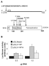
Figure 1
Figure 1
Characterization of mutation c-57-64delCACGCGGT;c-55G>C in the 5′ UTR of HNF1A. (A) Location of mutation c-57-64delCACGCGGT;c-55G>C in the 5′-UTR of the HNF1A gene and missense mutations in the HNF-1α protein. The HNF1A cDNA (upper panel) and the functional domains of HNF-1α protein (lower panel) are schematically represented. In the protein, amino acids 1–33 contain the dimerization domain (DD). DNA binding domain, including POUS and POUH homeodomains, is located between amino acids 100–281. The transactivation domain is spanning the C-terminal half part of the protein. The positions of the mutations analyzed are indicated. (B) Transcription of HNF1A is impaired by the c-57-64delCACGCGGT;c-55G>C mutation. Min6 cells, grown on six-well culture dishes, were cotransfected with the indicated amount of plasmids pGL3basic, pGL3-1AP and pGL3-1APm and 250 ng pCMVβ. Cells were harvested 24 h after transfection and assayed for luciferase and β-galactosidase activities. Luciferase activities, normalized to β-galactosidase, were relative to pGL3basic activity (arbitrarily given as 1). Data represent means ± SEM of four experiments done in duplicate. *P ≤ 0.05.
Figure 2
Figure 2
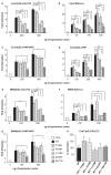
Transcriptional activity of mutants mHNF1(V133M), mHNF1(T196A), mHNF1(E235E), mHNF1(R271W) and mHNF1(P378R). Cos7 (A–D) and Min6 (E–G) cells were cotransfected with 250 of the reporter constructs pGL3-GLUT2, β28-Luc, pGL3-HNF4AP2 or pGL3-RIP; 200 ng pCMVβ; and the indicated amounts of wild-type or mutant HNF1A expression vectors, which were defined to obtain a sufficiently strong response with the wild-type (at least a two-fold activation) but avoid saturation of the system. Cells were harvested 24 h after transfection and assayed for luciferase and β-galactosidase activities. Luciferase activity was normalized by the β-galactosidase activity of the internal transfection efficiency control pCMVβ. For a typical experiment, β-galactosidase activity was 1.8 ± 0.1 (mean ± SEM) optical density (OD)420 units/[min • mg total protein]. Normalized luciferase values obtained from cells cotransfected with HNF1A expression vectors are shown as fold activation relative to the controls lacking any HNF1A expression vector, which were set as 1. (H) Cos7 cells were cotransfected with 200 ng pGL3-GLUT2, 200 ng pCMVβ and 100 ng wild-type pcDNA3-mHNF1A expression vector alone or together with 100 ng of each mutant expression vector, as indicated. Normalized luciferase values obtained from cells transfected with wild-type HNF-1α alone are set as 100%. All experiments were performed in duplicate and repeated at least 3× with different DNA preparations. The Student t test was used to compare the mean relative fold activation values between groups. Error bars indicate SEM. *P ≤ 0.05; **P ≤ 0.005. Note that human residue Pro379 corresponds to mouse Pro378.
Figure 3
Figure 3
(A) Schematic representation of the GST-fusion proteins containing mouse HNF-1α amino acids 1–281. DD, dimerization domain. DBD, DNA binding domain. Numbers indicate amino acid positions of the HNF-1α protein. (B) Binding site titration experiments using GST-fusion proteins GST-1α(1–281) and radiolabeled probe 1A. A total of 2 ng purified GST-1α(1–281) proteins were incubated with a varying amount of radiolabeled oligonucleotide (0.042, 0.084, 0.168, 0.336, 0.420, 0.672, 1.34, 2.68, 5.36 and 10.2 nmol/L). GST-1α(1–281)/DNA complexes were assayed by EMSA and quantified in a phosphorimager. Relative binding affinities (Ka ) and maximal binding (Bmax ) for the mutant and wild-type GST-1α(1–281) proteins were calculated by using the Scatchard plot. Data indicate mean ± SEM. Ka is given in nmol/L; Bmax is given in pmol DNA bound μg protein. n, Number of independent experiments. *P ≤ 0.05.
Figure 4
Figure 4
A Western blot analysis of nuclear extracts from Cos7 cells transfected with wild-type and HNF-1α variants. The 60-mm dishes of confluent cells were transfected with 10 μg of expression vectors using a modified calcium phosphate precipitation procedure (16). The 10 μL of nuclear extracts were subjected to 12% SDS-PAGE, followed by Western blotting analysis using the polyclonal anti–HNF-1α antiserum (C-19; Santa Cruz). mock, Cos7 cells transfected with no expression vector. (B) EMSA analysis of HNF-1α DNA binding activity. The 1 μL of nuclear extracts from Cos7 cells transfected with the expression vectors shown in (A) were incubated with 0.42 nmol/L 32P-labeled 1A or 4AP2 probes, as indicated. Lane 0, free probe; lane 1, band shift; lane 2, competition using a 25-fold excess of cold probe; lane 3, supershift using the polyclonal anti–HNF-1α antibody C-19. A representative experiment is shown.

References

    1. Murphy R, Ellard S, Hattersley AT. Clinical implications of a molecular genetic classification of monogenic beta-cell diabetes. Nat Clin Pract Endocrinol Metab. 2008;4:200–13. - PubMed
    1. Mendel DB, Crabtree GR. HNF-1, a member of a novel class of dimerizing homeodomain proteins. J Biol Chem. 1991;266:677–80. - PubMed
    1. Wang H, Maechler P, Hagenfeldt KA, Wollheim CB. Dominant-negative suppression of HNF-1alpha function results in defective insulin gene transcription and impaired metabolism-secretion coupling in a pancreatic beta-cell line. EMBO J. 1998;17:6701–13. - PMC - PubMed
    1. Shih DQ, et al. Loss of HNF-1alpha function in mice leads to abnormal expression of genes involved in pancreatic islet development and metabolism. Diabetes. 2001;50:2472–80. - PubMed
    1. Boj SF, Parrizas M, Maestro MA, Ferrer J. A transcription factor regulatory circuit in differentiated pancreatic cells. Proc Natl Acad Sci U S A. 2001;98:14481–6. - PMC - PubMed

Publication types

MeSH terms

Substances