. 2010 Dec 20:11:721.
doi: 10.1186/1471-2164-11-721.

The vertebrate makorin ubiquitin ligase gene family has been shaped by large-scale duplication and retroposition from an ancestral gonad-specific, maternal-effect gene

Affiliations

The vertebrate makorin ubiquitin ligase gene family has been shaped by large-scale duplication and retroposition from an ancestral gonad-specific, maternal-effect gene

Astrid Böhne et al. BMC Genomics. .

Abstract

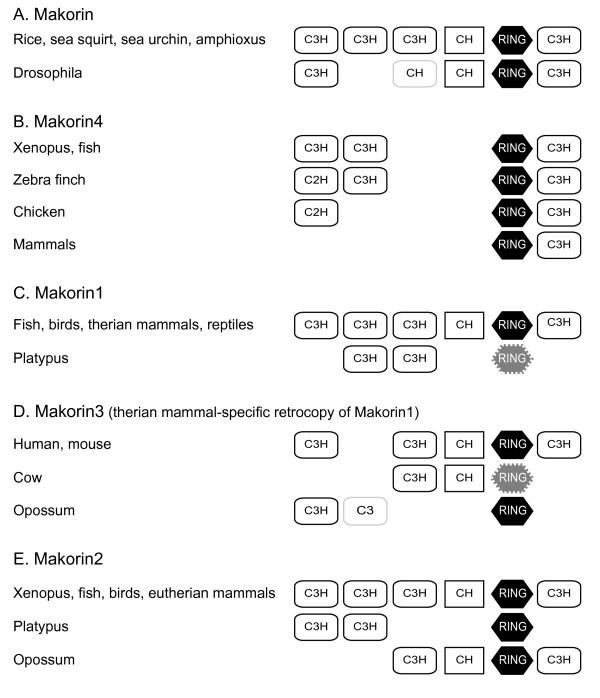
Background: Members of the makorin (mkrn) gene family encode RING/C3H zinc finger proteins with U3 ubiquitin ligase activity. Although these proteins have been described in a variety of eukaryotes such as plants, fungi, invertebrates and vertebrates including human, almost nothing is known about their structural and functional evolution.

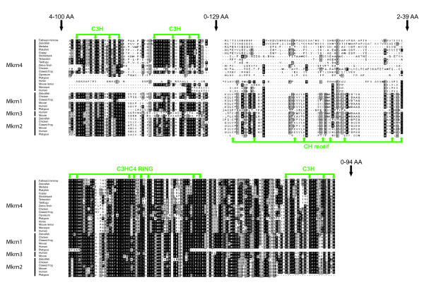
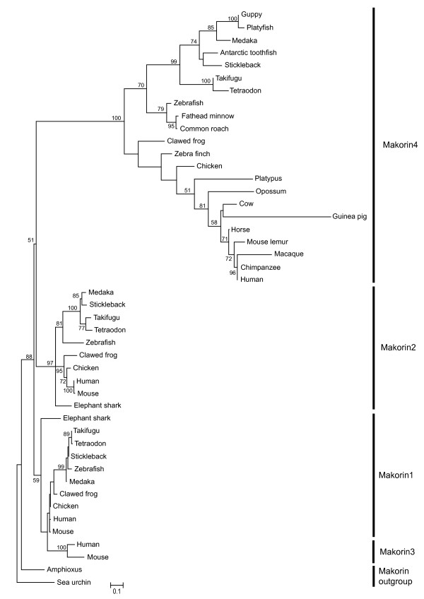
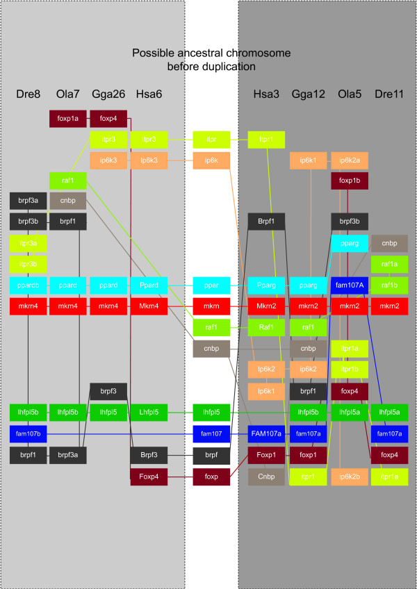
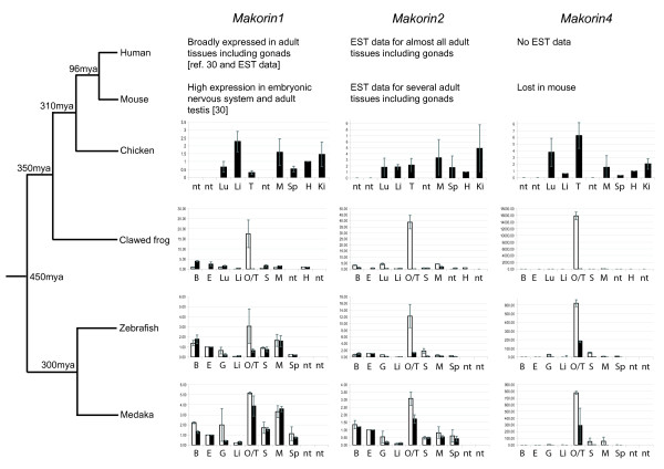
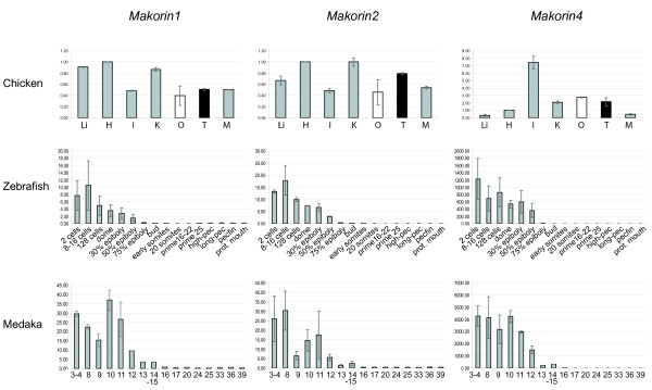
Results: Via partial sequencing of a testis cDNA library from the poeciliid fish Xiphophorus maculatus, we have identified a new member of the makorin gene family, that we called mkrn4. In addition to the already described mkrn1 and mkrn2, mkrn4 is the third example of a makorin gene present in both tetrapods and ray-finned fish. However, this gene was not detected in mouse and rat, suggesting its loss in the lineage leading to rodent murids. Mkrn2 and mkrn4 are located in large ancient duplicated regions in tetrapod and fish genomes, suggesting the possible involvement of ancestral vertebrate-specific genome duplication in the formation of these genes. Intriguingly, many mkrn1 and mkrn2 intronless retrocopies have been detected in mammals but not in other vertebrates, most of them corresponding to pseudogenes. The nature and number of zinc fingers were found to be conserved in Mkrn1 and Mkrn2 but much more variable in Mkrn4, with lineage-specific differences. RT-qPCR analysis demonstrated a highly gonad-biased expression pattern for makorin genes in medaka and zebrafish (ray-finned fishes) and amphibians, but a strong relaxation of this specificity in birds and mammals. All three mkrn genes were maternally expressed before zygotic genome activation in both medaka and zebrafish early embryos.

Conclusion: Our analysis demonstrates that the makorin gene family has evolved through large-scale duplication and subsequent lineage-specific retroposition-mediated duplications in vertebrates. From the three major vertebrate mkrn genes, mkrn4 shows the highest evolutionary dynamics, with lineage-specific loss of zinc fingers and even complete gene elimination from certain groups of vertebrates. Comparative expression analysis strongly suggests that the ancestral E3 ubiquitin ligase function of the single copy mkrn gene before duplication in vertebrates was gonad-specific, with maternal expression in early embryos.


Figures

Figure 1
Figure 1
Zinc finger domains in Mkrn4 and other vertebrate Makorin proteins. Green arrows show conserved cysteine and histidine residues in zinc finger motifs. Minimal/maximal numbers of amino-acid residues between motifs are indicated over black arrows. Black columns indicate identical amino-acids, grey columns similar residues.
Figure 2
Figure 2
Molecular phylogeny of the Makorin family. The tree was constructed using maximum likelihood analysis with 1000 bootstrap replicates on a 131 amino acid MUSCLE alignment of the RING domain and the C-terminal C3H zinc finger. The tree was rooted using the sequence of the sea urchin. Accession numbers of proteins used in the alignment are given in Additional file 1.
Figure 3
Figure 3
Mkrn2 and mkrn4 are included in large paralogons in bony vertebrates. Genes are indicated in colored boxes. Colored lines connect orthologs on one side with paralogs on the other side of the figure. Each grey panel contains a set of orthologous syntenic regions, the putative ancestral region before duplication is shown in the central white panel. Dre: Danio rerio (zebrafish); Gga: Gallus gallus (chicken); Hsa: Homo sapiens (human); Ola: Oryzias latipes (medaka fish). Accession numbers of genes presented in this figure are listed in Additional file 2.
Figure 4
Figure 4
Zinc finger domains in the Makorin protein family. Black hexagons indicate the RING domain, white rectangles the C3H zinc fingers. Dashed lines and lighter content indicate incomplete motifs due to sequence changes or loss. The structure of Mkrn2 for Xenopus is based on a X. laevis EST (EF626804) since the tropicalis sequence is incomplete and lacks the RING domain. Accession numbers are provided in Additional file 2.
Figure 5
Figure 5
RT-qPCR expression analysis of makorin genes in vertebrate adult tissues. Q-PCR values for each gene were normalized to expression levels of rpl7 using the 2-DDCT method [71]. Expression in eye (fish) or heart (chicken and frog) was set as a reference (value: 1); Data are presented as mean ± standard deviation of two independent quantitative real-time PCR experiments (average of two independent reverse transcription reactions, each tested with two PCR reactions). Black bars represent male tissues and organs, white bars female tissues and organs. B brain, E eye, G gills, H heart, I intestine, K kidneys, Lu lung, Li liver, M muscle, O ovary, S skin, Sp spleen, T testis, nt not tested.
Figure 6
Figure 6
RT-qPCR expression analysis of makorin genes in vertebrate embryos and embryonic tissues. Q-PCR values for each gene were normalized to expression levels of rpl7 using the 2-DDCT method [71]. Expression in adult eye (fish) (Fig. 5) or heart (chicken) was set as a reference (value: 1). Data are presented as mean ± standard deviation of two independent quantitative real-time PCR experiments (average of two independent reverse transcription reactions, each tested with two PCR reactions). Black bars represent male tissues and organs, white bars female tissues and organs. For grey bars, the sex of the donor(s) is undetermined (embryos prior to sexual differentiation) or both sexes have been mixed (frog samples). H heart, I intestine, K kidneys, Li liver, M muscle, O ovary, T testis. Embryonic stages are referred to as [53] for medaka and [54] for zebrafish, respectively.

Similar articles

Cited by

References

    1. Deshaies RJ, Joazeiro CA. RING domain E3 ubiquitin ligases. Annu Rev Biochem. 2009;78:399–434. doi: 10.1146/annurev.biochem.78.101807.093809. - DOI - PubMed
    1. Kim JH, Park SM, Kang MR, Oh SY, Lee TH, Muller MT, Chung IK. Ubiquitin ligase MKRN1 modulates telomere length homeostasis through a proteolysis of hTERT. Genes Dev. 2005;19:776–781. doi: 10.1101/gad.1289405. - DOI - PMC - PubMed
    1. Hall TM. Multiple modes of RNA recognition by zinc finger proteins. Curr Opin Struct Biol. 2005;15:367–373. doi: 10.1016/j.sbi.2005.04.004. - DOI - PubMed
    1. Gray TA, Hernandez L, Carey AH, Schaldach MA, Smithwick MJ, Rus K, Marshall Graves JA, Stewart CL, Nicholls RD. The ancient source of a distinct gene family encoding proteins featuring RING and C(3)H zinc-finger motifs with abundant expression in developing brain and nervous system. Genomics. 2000;66:76–86. doi: 10.1006/geno.2000.6199. - DOI - PubMed
    1. Ko A, Lee EW, Yang MR, Yeh JY, Moon JS, Song J. Mkrn1 induces the degradation of West Nile virus capsid protein by functioning as an E3 ligase. J Virol. 2009;84:426–436. doi: 10.1128/JVI.00725-09. - DOI - PMC - PubMed

Publication types

MeSH terms

LinkOut - more resources