. 2010 Dec 20:9:316.
doi: 10.1186/1476-4598-9-316.

A critical role for lymphatic endothelial heparan sulfate in lymph node metastasis

Affiliations

A critical role for lymphatic endothelial heparan sulfate in lymph node metastasis

Xin Yin et al. Mol Cancer. .

Abstract

Background: Lymph node metastasis constitutes a key event in tumor progression. The molecular control of this process is poorly understood. Heparan sulfate is a linear polysaccharide consisting of unique sulfate-modified disaccharide repeats that allow the glycan to bind a variety of proteins, including chemokines. While some chemokines may drive lymphatic trafficking of tumor cells, the functional and genetic importance of heparan sulfate as a possible mediator of chemokine actions in lymphatic metastasis has not been reported.

Results: We applied a loss-of-function genetic approach employing lymphatic endothelial conditional mutations in heparan sulfate biosynthesis to study the effects on tumor-lymphatic trafficking and lymph node metastasis. Lymphatic endothelial deficiency in N-deacetylase/N-sulfotransferase-1 (Ndst1), a key enzyme involved in sulfating nascent heparan sulfate chains, resulted in altered lymph node metastasis in tumor-bearing gene targeted mice. This occurred in mice harboring either a pan-endothelial Ndst1 mutation or an inducible lymphatic-endothelial specific mutation in Ndst1. In addition to a marked reduction in tumor metastases to the regional lymph nodes in mutant mice, specific immuno-localization of CCL21, a heparin-binding chemokine known to regulate leukocyte and possibly tumor-cell traffic, showed a marked reduction in its ability to associate with tumor cells in mutant lymph nodes. In vitro modified chemotaxis studies targeting heparan sulfate biosynthesis in lymphatic endothelial cells revealed that heparan sulfate secreted by lymphatic endothelium is required for CCL21-dependent directional migration of murine as well as human lung carcinoma cells toward the targeted lymphatic endothelium. Lymphatic heparan sulfate was also required for binding of CCL21 to its receptor CCR7 on tumor cells as well as the activation of migration signaling pathways in tumor cells exposed to lymphatic conditioned medium. Finally, lymphatic cell-surface heparan sulfate facilitated receptor-dependent binding and concentration of CCL21 on the lymphatic endothelium, thereby serving as a mechanism to generate lymphatic chemokine gradients.

Conclusions: This work demonstrates the genetic importance of host lymphatic heparan sulfate in mediating chemokine dependent tumor-cell traffic in the lymphatic microenvironment. The impact on chemokine dependent lymphatic metastasis may guide novel therapeutic strategies.


Figures
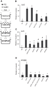
Figure 1
Figure 1
Invasion of tumor cells toward matrix-embedded lymphatic endothelial cells depends on lymphatic heparan sulfate. A. Schematic representation of a modified transwell collagen invasion assay. TC, tumor cells; hLEC, primay human lung lymphatic endothelial cells; Col, type I collagen gel. B and C. Invasion of Lewis lung carcinoma cells (LLC) toward collagen gel containing either no cells ("NC") or hLEC treated as indicated was quantified and normalized to NC. αCCL21, αCCR7 or αCCL5, blocking antibodies to CCL21, CCR7 or CCL5, respectively; H'ase, hLEC pre-treated with heparinase; siDS, hLEC transfected with control (scrambled duplex) RNA; siNdst1, siXylT2, siHs2st or siHs3st1, hLEC transfected with siRNA targeting corresponding HS biosynthetic enzymes. D. Invasion of human lung adenocarcinoma cells (H1650) toward collagen gel containing either no cells ("NC") or hLEC treated as indicated was quantified and normalized to NC. *P < 0.01, #P < 0.05, as compared to hLEC in B and siDS in C and D.
Figure 2
Figure 2
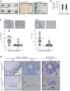
Genetic targeting of pan-endothelial heparan sulfate biosynthesis impairs metastasis of carcinoma to regional lymph nodes. LLC tumor cells were injected subcutaneously into the left caudal/medial inguinal region of Ndst1f/fTekCre+ mutant mice, which bear a pan-endothelial mutation in the major HS sulfating enzyme Ndst1, and their wildtype Cre- littermates as controls. After 14 days, the left subiliac lymph node (draining the primary tumor) from each mouse was isolated. A. Metastaic tumor cells in the lymph nodes (LN) were detected using anti-pan-keratin antibody (blue stain) on LN tissue sections imaged under 100× magnification, and quantified (NIH Image-J) to determine net pixel intensity for each LN. Values were normalized to the mean pixel value/LN among control (Ndst1f/fTekCre-) littermates. Graph is shown below; *P = 0.016, for difference in mean values (red bars) for mutant (N = 8) vs control (N = 6) mice. B. Representative LN tissue sections from tumor-bearing mutant (bottom row of photomicrographs) vs wildtype (Cre-) controls (middle row) were co-stained with CCL21 (blue) and LYVE1 (brown) (left two panels) or pan-keratin (blue, right panel). The top row of photomicrographs shows a representative LN section from a non-challenged (tumor free) control mouse. Scale bars, 100 μm for 100× and 50 μm for 400× magnification photomicrographs.
Figure 3
Figure 3
Migration of Lewis lung carcinoma cells toward lymphatic endothelial cells depends on lymphatic heparan sulfate. A. Schematic representation of a modified transwell chemotaxis assay, wherein liquid medium separates migrating LLC tumor cells that initiate in the upper well and migrate toward lower wells that contain either no cells or hLEC monolayers treated under various conditions. B and C. Transwell migration of tumor cells into lower wells plated with either no cells as a negative control ("NC") or hLEC monolayers treated as indicated was quantified, and plotted for each condition as the mean -fold response over the value for NC (and normalized to the NC value). Conditions in B refer to the addition of specific blocking antibodies or pre-treatment of the hLEC with heparinase (H'ase); and conditions in C refer to treatment of the hLEC with siRNA targeting the indicated HS biosynthetic enzymes. *P < 0.01, #P < 0.05, as compared to hLEC group in B and siDS group in C. D. Graphs quantifying transwell migration of either LLC (upper graph) or H1650 (lower graph) lung carcinoma cells into lower wells plated with either no cells (NC), control hLEC (siDS), or siXylT2 targeted hLEC. The condition "siXylT2+HS" (far right) refers to addition of heparan sulfate purified from cultured control hLEC into the lower-well medium during tumor cell migration toward siXylT2 targeted hLEC. Fractions (0.04 and 0.2) of the total HS needed for rescue were used in separate wells to test dose-response. #P < 0.05, *P < 0.01, **P < 0.001 for histogram comparisons indicated by horizontal bars.
Figure 4
Figure 4
Lymphatic-secreted HS activates tumor-cell migration signaling pathways in trans. A. Serum-free basal endothelial growth medium or conditioned medium (CM) harvested from hLEC treated as indicated was applied to cultured LLC for 10 min. The LLC were lysed and the indicated target proteins were detected by Western immunoblotting. A representative gel image (top) as well as normalized densitometric quantification (below) from three independent experiments is shown. Relative protein expression was calculated as the densitometric ratio of phosphorylated protein to that of corresponding total protein, and normalized to the basal value. *P < 0.05, as compared to LLC treated with CM from control hLEC (CM-siDS). B. In separate experiments, the effects of blocking CCL21 (siDS + αCCL21) or treatments that alter HS biosynthesis (siNdst1 or siXylT2) on CM-mediated Erk1/2 phosphorylation were examined.
Figure 5
Figure 5
Heparan sulfate in the lymphatic conditioned medium is required for CCL21 binding to tumor cells. Conditioned medium (CM) from hLEC transfected with either control (scrambled duplex) RNA (siDS) or siRNA targeting the indicated HS biosynthetic enzymes was harvested and applied to LLCs (A) or H1650 (B) cytospin samples, respectively. Binding of CCL21 in the CM to CCR7 on LLC was detected by proximity ligation assay (PLA). Representative merged images showing PLA signal (red) and nuclear DAPI stain (blue) were taken by fluorescence microscopy (400 ×)(A and B photomicrographs; Scale bar, 50 μm.) PLA signal from each field was quantified and indexed to total nuclear area within the same field (A and B graphs). At least 5 random fields from each group were included for analysis. Mean data was normalized to control signal (CM-siDS). *P < 0.05, #P < 0.01, as compared to CM-siDS group. C. For each hLEC siRNA treatment condition, CCL21 in both the cell lysate (left) and CM were detected by Western immunoblot analysis. Tubulin was probed as a loading control. D. HS was purified from CM of siDS- or siNdst1-transfected hLEC and the sulfation status examined by disaccharide analysis using liquid chromatography/mass spectrometry.
Figure 6
Figure 6
Lymphatic endothelial heparan sulfate is required for receptor-dependent display of CCL21 on the lymphatic surface. A. Binding of human CCL21 (hCCL21) to heparin was examined using heparin affinity chromatography followed by silver staining of salt-eluted fractions on SDS-PAGE (upper panel). "Pre," recombinant human CCL21 sample directly loaded onto the silver-stained gel; "FT," flow-through from the column. Basic fibroblast growth factor (FGF-2) was run as a positive control (lower panel). B. hLEC were treated as indicated and the binding of CCL21 to hLEC was examined by immunofluorescence (IF) using anti-CCL21 antibody. hLEC, untreated cells; H'ase, Heparin: hLEC pre-treated with heparinase or heparin, respectively; H'ase+CCL21, Heparin+CCL21: hLEC pre-treated with heparinase or heparin, respectively, followed by addition of exogenous human recombinant CCL21 (+CCL21). Representative merged images showing CCL21 signal (green) and DAPI nuclear stain (blue) were taken under 100× magnification. (Scale bar, 100 μm.) C. hLEC were transfected with either control RNA (siDS) or siRNA targeting HS biosynthetic enzymes Ndst1 or XylT2. Binding CCL21 to CCR7 on hLEC was detected by proximity ligation assay (PLA). Representative merged images showing PLA signal (red) and nuclear DAPI stain (blue), imaged using fluorescence microscopy (400×; Scale bar, 50 μm). PLA signal from each field was quantified and indexed to total nuclear area within the same field, and mean values for each condition were graphed (bottom). At least 5 random fields from each group were included for analysis. Mean data was normalized to control signal (siDS). *P < 0.01, as compared to control.
Figure 7
Figure 7
Genetic alteration of lymphatic endothelial heparan sulfate biosynthesis impairs tumor metastasis to regional lymph nodes. A. Cre reporter testing in the inducible Prox1+/CreERT2 model: Bilateral subiliac lymph nodes (LN) were isolated from Prox1+/CreERT2Rosa26R reporter mice ("Prox1+/CreERT2", lower panel) or their Prox1-/CreERT2Rosa26R littermates ("Prox1-/CreERT2", upper panel) after intraperitoneal injection of Tamoxifen for 5 consecutive days; and stained with X-Gal (showing as deep blue stain in Cre positive lymph nodes). Right panels: subiliac LNs from A were sectioned and stained for LYVE-1 (brown) and X-Gal (blue); and imaged under 400 × magnification (Scale bar, 50 μm). For tumor establishment, LLC tumor cells were injected subcutaneously into the left caudal-medial inguinal region of Ndst1f/fProx1+/CreERT2 mice (N = 10) and their Ndst1f/fProx1-/CreERT2 wildtype littermates (N = 10) following a 5-day tamoxifen dosing schedule. Following 14 days of primary tumor growth, the left subiliac regional LN from each mouse was isolated. B. Comparison of primary tumor volumes from Cre postive vs Cre negative mice at time of experiment termination and lymph node harvesting. C. Representative photomicrographs of pan-keratin stained LN histologic sections are shown above plotted data on the left, which shows quantified pan-keratin signal per LN for each mouse. Graph to the right shows plotted data for degree of CCL21 immunoreactive signal per LN (and representative photomicrographs of CCL21 staining from Cre positive vs Cre negative animals above the graph). LN staining signal from both groups was imaged under 100× magnification, quantified as pixel values per LN using NIH Image J software, and normalized to the average pixel value/LN in Ndst1f/fProx1-/CreERT2 control group. *P < 0.05, #P < 0.01, for comparison of mean mutant vs control group values. D. LNs from both groups were co-stained with CCL21 (blue) and LYVE1 (brown) (left two panels) or pan-keratin alone as a marker for metastatic tumor cells (blue signal, right panel). Scale bars, 100 μm for 100× and 50 μm for 400× magnification.
Figure 8
Figure 8
Summary of mechanistic considerations: Role of lymphatic endothelial heparan sulfate in chemokine-mediated lymph node metastasis. A. In peripheral lymphatic vessels (e.g., lymphatic vascular bed of a primary tumor), HS-binding chemokines produced by the lymphatic endothelium such as CCL21 may "cluster" on lymphatic-bound as well as secreted HS proteoglycans. In the setting of intra-lymphatic flow (thin arrows), spatial gradients of the chemokine scaffolded on HS chains in the extracellular matrix and lymphatic endothelium may facilitate tumor-cell migration into lymphatic vessels. This may occur in coordination with other adhesion systems such as integrins or selectins. Clustering of chemokines (as dimers in this example) on HS in the peri-lymphatic extracellular matrix may also be critical for receptor activation (e.g., CCR7) on migrating tumor cells. Targeting HS biosynthesis may thus alter chemokine-receptor interactions (large black arrow, mechanism "1") as well as chemokine gradients (large grey arrow, mechanism "2"). B. In the lymph node, similar principles may apply; however, the direction of trans-lymphatic flow and chemokine gradient are reversed, facilitating extra-vasation (colonization) of trafficking tumor cells expressing cognate chemokine receptors. Interfering with the biosynthesis of lymphatic heparan sulfate (e.g., genetically targeting the sulfation of HS through lymphatic endothelial Ndst1 mutation) may thus abrogate lymph node metastasis by altering such co-receptor and gradient-mediating functions served by the glycan.

Similar articles

Cited by

References

    1. Detterbeck FC, Boffa DJ, Tanoue LT. The new lung cancer staging system. Chest. 2009;136(1):260–271. doi: 10.1378/chest.08-0978. - DOI - PubMed
    1. Effenberger KE, Sienel W, Pantel K. Lymph node micrometastases in lung cancer. Cancer Treat Res. 2007;135:167–175. full_text. - PubMed
    1. Perez-Cardona JH, Ordonez NG, Fossella FV. Lymph node micrometastases in non small-cell lung cancer: clinical applications. Clin Lung Cancer. 2000;2(2):116–120. doi: 10.3816/CLC.2000.n.023. - DOI - PubMed
    1. Alitalo K, Tammela T, Petrova TV. Lymphangiogenesis in development and human disease. Nature. 2005;438(7070):946–953. doi: 10.1038/nature04480. - DOI - PubMed
    1. Cao Y. Opinion: emerging mechanisms of tumour lymphangiogenesis and lymphatic metastasis. Nat Rev Cancer. 2005;5(9):735–743. doi: 10.1038/nrc1693. - DOI - PubMed

Publication types

MeSH terms