. 2010 Dec 22;40(6):1016-23.
doi: 10.1016/j.molcel.2010.11.024.

TDRD3 is an effector molecule for arginine-methylated histone marks

Affiliations

TDRD3 is an effector molecule for arginine-methylated histone marks

Yanzhong Yang et al. Mol Cell. .

Abstract

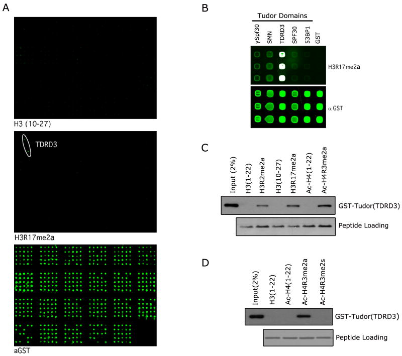
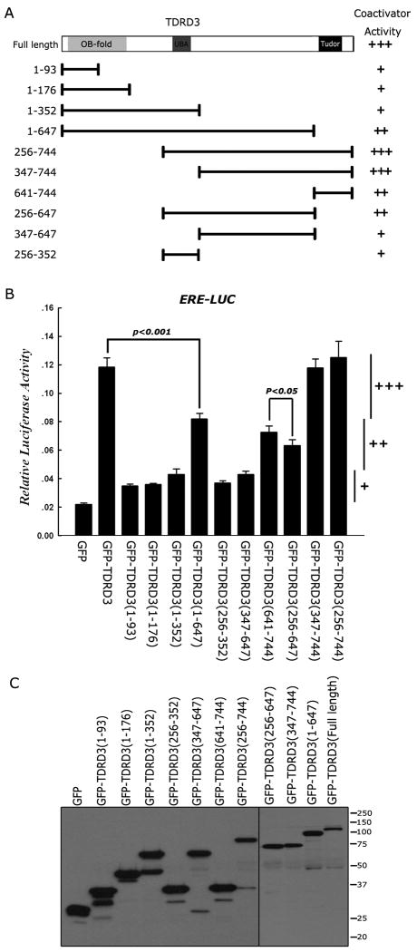
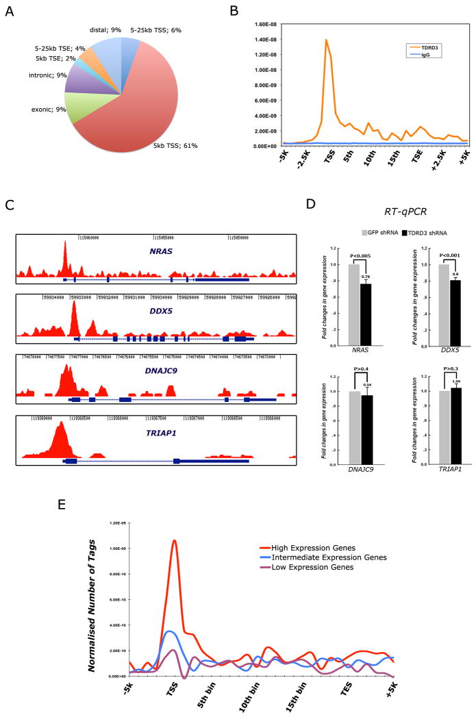
Specific sites of histone tail methylation are associated with transcriptional activity at gene loci. These methyl marks are interpreted by effector molecules, which harbor protein domains that bind the methylated motifs and facilitate either active or inactive states of transcription. CARM1 and PRMT1 are transcriptional coactivators that deposit H3R17me2a and H4R3me2a marks, respectively. We used a protein domain microarray approach to identify the Tudor domain-containing protein TDRD3 as a "reader" of these marks. Importantly, TDRD3 itself is a transcriptional coactivator. This coactivator activity requires an intact Tudor domain. TDRD3 is recruited to an estrogen-responsive element in a CARM1-dependent manner. Furthermore, ChIP-seq analysis of TDRD3 reveals that it is predominantly localized to transcriptional start sites. Thus, TDRD3 is an effector molecule that promotes transcription by binding methylarginine marks on histone tails.


Figures

Figure 1
Figure 1
The tudor domain of TDRD3 binds arginine methylation marks on histone tails. (A) The CADOR array was probed with a peptide harboring the CARM1 methyl-mark H3R17me2a. Binding was observed on two spots representing the tudor domain of TDRD3 (middle panel). The unmodified peptide displays no binding under the same conditions (upper panel). The array complexity is revealed by probing with an anti-GST antibody (bottom panel). The array is composed of GST fusion proteins that carry protein domains usually found in chromatin associated proteins (for key see Figure S1). Proteins are arrayed in duplicate. (B) A focused array was probed using tudor domains that have been reported to bind methylarginine motifs (ySPF30, SMN, TDRD3 & SPF30) and the methyllysine binding tudor domain of 53BP1. The H3R17me2a peptide binds most strongly to the tudor domain of TDRD3. (C) The tudor domain of TDRD3 binds a number of methylarginine marks found on histone tails. Pull-down experiments were performed with H3R2me2a, H3R17me2a and Ac-H4R3me2a peptides. The observed pull-down efficiency is as follows: Ac-H4R3me2a > H3R17me2a > H3R2me2a. (D) TDRD3 tudor binds the asymmetric, but not the symmetric, dimethylarginine mark on H4R3. Pull-down experiments were performed with Ac-H4R3me2s and Ac-H4R3me2a peptides. A specificity switch is observed with the tudor domain of TDRD3 which binds more strongly to the Ac-H4R3me2a active mark (see also Figure S1).
Figure 2
Figure 2
TDRD3 is a transcriptional coactivator that is recruited to the pS2 promoter in a CARM1 dependent manner. (A) An ERE-Luc reporter assay was performed in MCF7 cells by co-transfection of GFP-SPF30, GFP-SMN and GFP-TDRD3 expression vectors. (B) ERE-Luc reporter activity is reduced in MCF7 cells with shRNA-mediated knockdown of endogenous TDRD3. Error bars represent standard deviation calculated from triplicate luciferase assays. (C) and (D) The recruitment of TDRD3 to the promoter of the estrogen-responsive pS2 promoter was evaluated by ChIP/ReChIP. MCF7 cells were cultured in charcoal-striped serum for three days and then treated with estradiol (E2+) for 45 min before ChIP/ReChIP experiments were performed with indicated antibodies. Immunoprecipitated DNA was analyzed in triplicates by qPCR with primers flanking the pS2 promoter region. Mean values were expressed as the fold change of E2+ versus E2-. E2- was represented as 1. (E) TDRD3 knockdown results in a reduction of the pS2 expression level. Hormone starved MCF7 cells were transfacted with shRNA against either GFP or TDRD3, respectively. Cells were either untreated or treated with E2 for 30 min. Total RNA was analyzed by RT-qPCR for pS2 expression and normalized for GAPDH. Error bars represent standard deviation calculated from triplicate qPCR reactions (see also Figure S2).
Figure 3
Figure 3
TDRD3 requires its tudor domain for optimal coactivator activity. (A) A series of GFP-fusion deletion constructs was generated. The region fused to GFP is graphically depicted. (B) An ERE-Luc reporter assay was performed in MCF7 cells by cotransfection of GFP-fusion deletion expression vectors. Relative activity is scored with +, ++ or +++. These scores are also shown in (A) to facilitate the interpretation of the data. Student t-test was performed for the indicated pairs. (C) The deletion construct series is expressed at roughly the same level. Western analysis was performed with αGFP. Error bars represent standard deviation calculated from triplicate luciferase assays.
Figure 4
Figure 4
TDRD3 is predominantly recruited to transcriptional start sites (TSS) and promoters of genes. (A) TDRD3 ChIP-seq analysis was performed using MCF7 cells. A pie chart is used to illustrate the genomic regions that are enriched for TDRD3 association. TDRD3 is predominantly coupled to promoter regions, including the TSS. (B) The distribution of TDRD3 was normalized across all genes in which peaks were identified, and its localization spikes over promoters, and displays some enrichment over the gene body. The TSS and TSE (transcriptional end) are shown. (C) Raw reads of the ChIP-seq tracings for four genes – NRAS, DDX5, DNAJC9 and TRIAP1 – demonstrating the enrichment of TDRD3 at promoters. (D) MCF7 cells were transfected with shRNA against GFP or TDRD3. Total RNA was extracted and RT-qPCR was performed to detect the level of gene expression at these four loci. Error bars represent standard deviation calculated from triplicate qPCR reactions. (E) Correlations between the level of gene expression in MCF7 cells and the occurrence of TDRD3 peaks at promoters. Gene expression levels were gauged using Affymetrix arrays (see also Figure S3).

References

    1. An W, Kim J, Roeder RG. Ordered cooperative functions of PRMT1, p300, and CARM1 in transcriptional activation by p53. Cell. 2004;117:735–748. - PubMed
    1. Bannister AJ, Zegerman P, Partridge JF, Miska EA, Thomas JO, Allshire RC, Kouzarides T. Selective recognition of methylated lysine 9 on histone H3 by the HP1 chromo domain. Nature. 2001;410:120–124. - PubMed
    1. Bedford MT, Clarke SG. Protein arginine methylation in mammals: who, what, and why. Mol Cell. 2009;33:1–13. - PMC - PubMed
    1. Chen C, Jin J, James DA, Adams-Cioaba MA, Park JG, Guo Y, Tenaglia E, Xu C, Gish G, Min J, et al. Mouse Piwi interactome identifies binding mechanism of Tdrkh Tudor domain to arginine methylated Miwi. Proc Natl Acad Sci U S A. 2009;106:20336–20341. - PMC - PubMed
    1. Chen D, Ma H, Hong H, Koh SS, Huang SM, Schurter BT, Aswad DW, Stallcup MR. Regulation of transcription by a protein methyltransferase. Science. 1999;284:2174–2177. - PubMed

Publication types

MeSH terms