Review
. 2010 Dec;2(12 Suppl 2):S253-68.
doi: 10.1016/j.pmrj.2010.10.015.

Assessment and modulation of neural plasticity in rehabilitation with transcranial magnetic stimulation

Affiliations
Review

Assessment and modulation of neural plasticity in rehabilitation with transcranial magnetic stimulation

Shahid Bashir et al. PM R. 2010 Dec.

Abstract

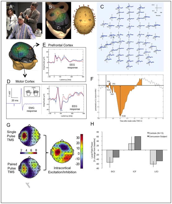
Despite intensive efforts to improve outcomes after acquired brain injury, functional recovery is often limited. One reason for this limitation is the challenge in assessing and guiding plasticity after brain injury. In this context, transcranial magnetic stimulation (TMS), a noninvasive tool of brain stimulation, could play a major role. TMS has been shown to be a reliable tool for measuring plastic changes in the motor cortex associated with interventions in the motor system, such as motor training and motor cortex stimulation. In addition, as illustrated by the experience in promoting recovery from stroke, TMS is a promising therapeutic tool to minimize motor, speech, cognitive, and mood deficits. In this review, we will focus on stroke to discuss how TMS can provide insights into the mechanisms of neurologic recovery and how it can be used for measurement and modulation of plasticity after an acquired brain insult.


Figures

FIGURE 1
FIGURE 1. Summary of TMS methodology
(A) The Nexstim TMS system is one of the available systems that provides neuronavigation and real-time display of induced current in the subject’s brain and targeted brain area (B), and enables fully integrated EMG (D) and EEG (C, D,E) recording. TMS-evoked EEG potentials (D, E) can be quantified using various measures of amplitude, timing and magnitude (F) and topographic distribution (G), allowing quantification of various measures of E/I balance. H demonstrates feasibility of the use of this techniques in the assessment of patients with acquired brain insults, in this case mild traumatic brain injury. It shows the average response to ICF, SICI and LICI for 13 control subjects and a pilot participant with a recent concussion. Controls show the expected suppression to SICI and LICI and enhancement in ICF. The TBI patient, shows less SICI and LICI, and more ICF than controls. Note that a subject with concussion shows reduced SICI and LICI, and increased ICF (as compared with 13 normal controls ± 95% confidence intervals). Navigated TMS utilizes individual structural MRI to enable online monitoring of the targeted cortical region, as well as coil position and orientation within and across sessions. The Nexstim system also calculates and displays the results of finite element models of the induced currents in each participant’s brain, and integrates a high-density EEG system. This provides trial by trial information about the induced electric field for every TMS pulse, the localization of the induced current maxima, all the TMS parameters, and the recorded EEG free of artifacts thanks to a pin-and-hold circuit. Such a system provides the reproducibility of the stimulation parameters that is essential for any comparative or longitudinal study. Critically, the calculation and display of the induced currents will allows to overcome the limitation of TMS targeting according to anatomical brain structures alone, since interindividual differences in structure–function relationships, particularly in individuals with brain pathology, may alter the induced currents and thus precision of the neuronavigation system.
FIGURE 2
FIGURE 2. TMS-EEG measures of cortical reactivity and plasticity
TMS-EEG measures of cortical reactivity (E/I balance) before (A) and after (B) continuous theta burst (cTBS) in order to assess cortical plasticity (LTD-like). EEG measures can be quantified in various ways (C, D). Single pulse TMS can induce TMS-evoked potentials (TEP) that can be measured by EEG. When applying TMS to the motor cortex, EMG will also be used to define motor threshold and silent period. TEPs will be characterized by N100 amplitude, P30-N45 peak-to-peak amplitude and Global Field Power. Paired pulse TMS delivered at different interpulse intervals can determine various aspects of intracortical inhibition and facilitation (SICI, ICF, LICI). Continuous theta burst stimulation (cTBS) can be applied as three pulses at 50 Hz repeated at 200 ms intervals (80% of active motor threshold intensity). Participants can receive a 47 s train of uninterrupted TBS (600 pulses). This paradigm causes suppression of post-stimulation MEPs in healthy subjects. The measures of intracortical E/I balance described above can be obtained before and then serially after cTBS (immediately after, T0, and then every 5 min following cTBS to track changes in amplitude over time). As an index of the duration of the cTBS-induced modulation of cortical response (index of plasticity), one can define for each participant the time point at which the average E/I measure returns within the 95% confidence interval of baseline and does not return to outside that interval on subsequent time point measures. E presents pilot data in controls and a patient with a recent concussion to illustrate the value of such measures in assessing the neurophysiologic impact of acquired brain injury. Control subjects show the expected suppression following cTBS and lasting approximately 30 min (LTD-like plasticity). However, the TBI subject shows a marked suppression of LTD-like plasticity, a return to baseline following cTBS after less than 5 min, and an apparent inversion of the modulatory effects of cTBS (potentiation instead of depression). Notably, such neurophysiologic effects are demonstrable despite absent clinical deficit. Contrasting the results of TMS on EEG before and after TBS will provide information about cortical plasticity (at the site of stimulation) but also about resulting network dynamic adaptation. Brain functional connectivity and inter-regional coordination can be directly estimated from EEGs. Analysis of network dynamics over time in patients with concussion will be compared against normal controls. For example, local, inter-region and global synchronization will be estimated, using 1) cross-correlation of EEG signals in the time domain, which will enable us to estimate temporal locking of these potentials; 2) cross-coherence in the frequency domain, assuming stationarity of the data in the analysis window; and 3) relative phase, obtained from phase time series corresponding to each EEG signal. Multivariate mixed effects regression models will be developed to compare measures of network synchronization and direct effects of stimulation across brain regions, time points, and study cohorts.

References

    1. Andlin-Sobocki P, Jonsson B, Wittchen HU, Olesen J. Cost of disorders of the brain in Europe. Eur J Neurol. 2005 Jun;12( Suppl 1):1–27. - PubMed
    1. Rossini PM, Calautti C, Pauri F, Baron JC. Post-stroke plastic reorganisation in the adult brain. Lancet Neurol. 2003 Aug;2(8):493–502. - PubMed
    1. Seitz RJ, Azari NP, Knorr U, Binkofski F, Herzog H, Freund HJ. The role of diaschisis in stroke recovery. Stroke. 1999 Sep;30(9):1844–1850. - PubMed
    1. Pascual-Leone A, Amedi A, Fregni F, Merabet LB. The plastic human brain cortex. Annu Rev Neurosci. 2005;28:377–401. - PubMed
    1. Dijkhuizen RM, Asahi M, Wu O, Rosen BR, Lo EH. Delayed rt-PA treatment in a rat embolic stroke model: diagnosis and prognosis of ischemic injury and hemorrhagic transformation with magnetic resonance imaging. J Cereb Blood Flow Metab. 2001 Aug;21(8):964–971. - PubMed

Publication types