. 2011 Jan 15;124(Pt 2):280-91.
doi: 10.1242/jcs.075390. Epub 2010 Dec 20.

PML isoforms I and II participate in PML-dependent restriction of HSV-1 replication

Affiliations

PML isoforms I and II participate in PML-dependent restriction of HSV-1 replication

Delphine Cuchet et al. J Cell Sci. .

Abstract

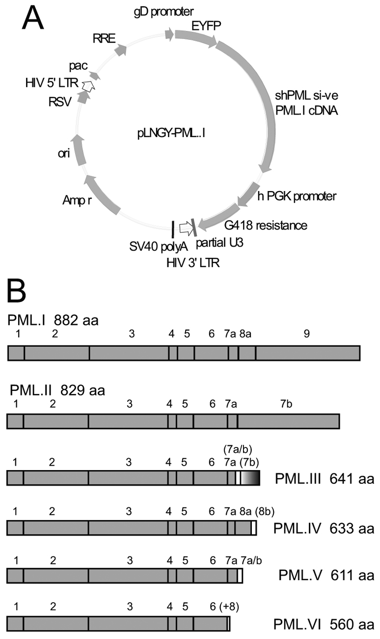
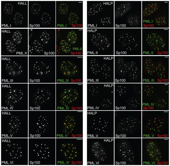
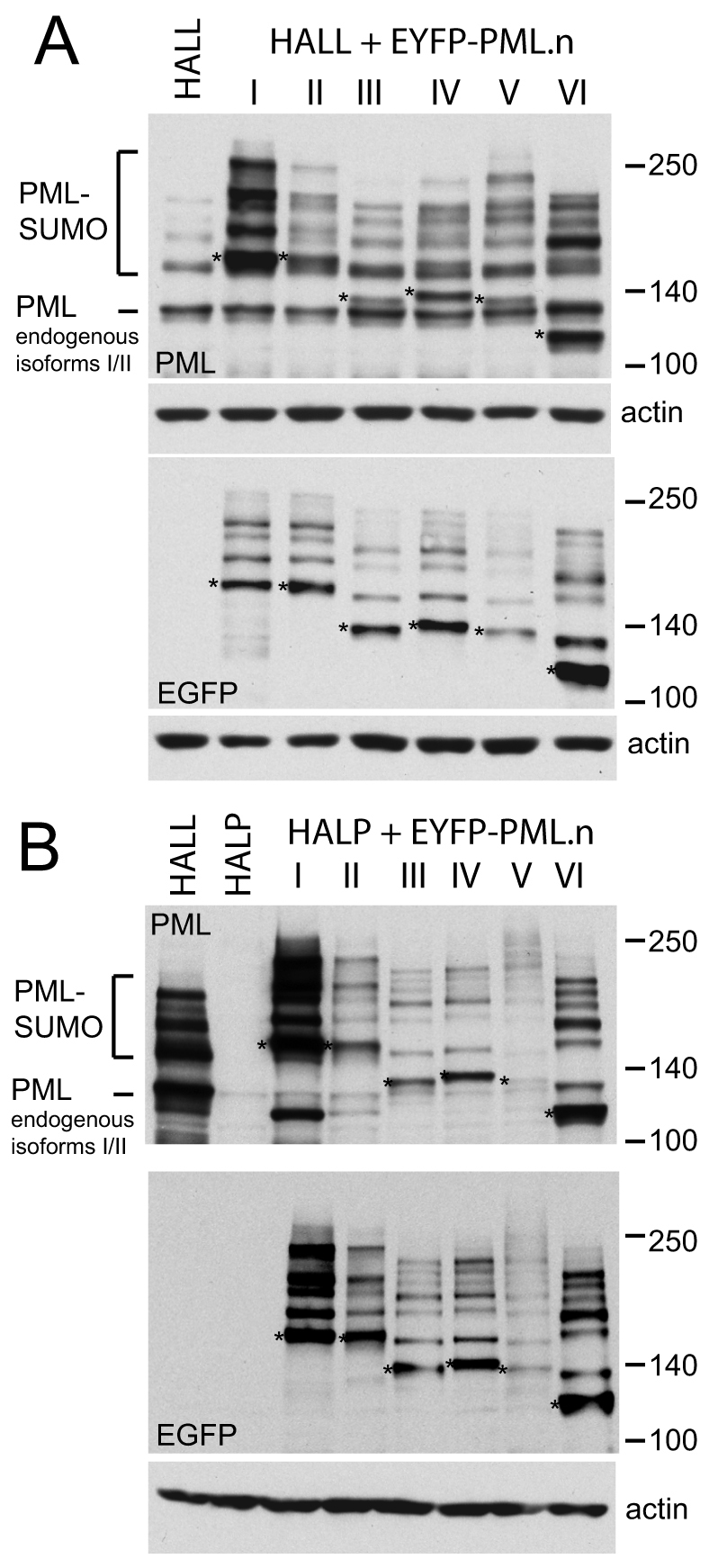
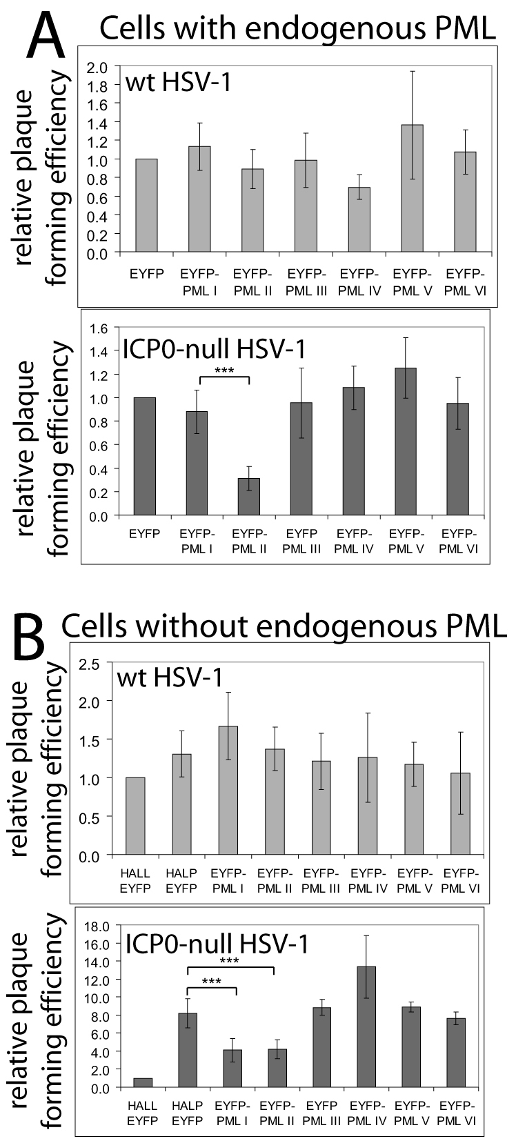
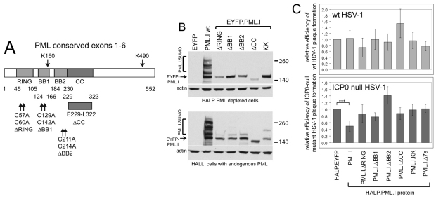
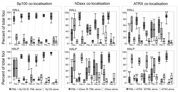
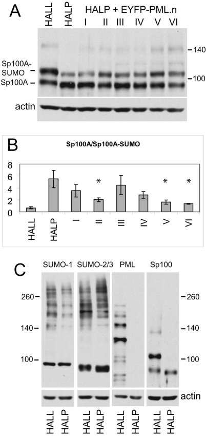
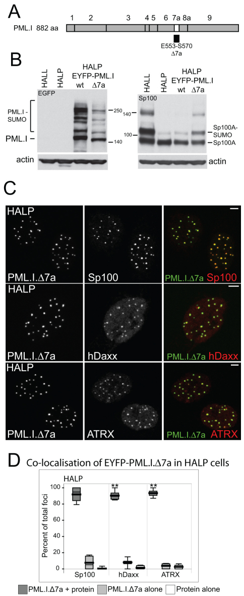
Intrinsic antiviral resistance mediated by constitutively expressed cellular proteins is one arm of defence against virus infection. Promyelocytic leukaemia nuclear bodies (PML-NBs, also known as ND10) contribute to host restriction of herpes simplex virus type 1 (HSV-1) replication via mechanisms that are counteracted by viral regulatory protein ICP0. ND10 assembly is dependent on PML, which comprises several different isoforms, and depletion of all PML isoforms decreases cellular resistance to ICP0-null mutant HSV-1. We report that individual expression of PML isoforms I and II partially reverses the increase in ICP0-null mutant HSV-1 plaque formation that occurs in PML-depleted cells. This activity of PML isoform I is dependent on SUMO modification, its SUMO interaction motif (SIM), and each element of its TRIM domain. Detailed analysis revealed that the punctate foci formed by individual PML isoforms differ subtly from normal ND10 in terms of composition and/or Sp100 modification. Surprisingly, deletion of the SIM motif from PML isoform I resulted in increased colocalisation with other major ND10 components in cells lacking endogenous PML. Our observations suggest that complete functionality of PML is dependent on isoform-specific C-terminal sequences acting in concert.


Figures

Fig. 1.
Fig. 1.
Map of the lentiviral vector used for PML expression and a summary of the PML isoforms studied. (A) Lentivirus plasmid vector pLNGY-PML.I. The key features of the lentivirus vector are noted: pac, HIV packaging sequence; RRE, REV response element; RSV, RSV promoter; ori, bacterial origin of replication; Amp r, ampicillin resistance; shPML1 si-ve, position of target sequence of shPML1 and relevant non-coding mutations (other labels are self-explanatory). (B) PML isoforms I to VI, noting the exons included in each isoform cDNA and the size of the translated product. The bracketed exon labels indicate the use of alternative reading frames compared to unbracketed exons of the same name. The (+8) label of PML.VI indicates that an additional eight residues follow exon 6 as a result of an alternative splicing event that deletes exon 7a.
Fig. 2.
Fig. 2.
Colocalisation of Sp100 and EYPF-tagged PML isoforms in HALL and HALP cells. Immunofluorescence staining of endogenous Sp100 in HALL (left-hand set of images) and HALP cells (right-hand set of images) expressing PML isoforms I to VI as EYFP fusion proteins (detected by EYFP autofluorescence). Images are representative of the majority phenotype in each cell line. The images are single plane projections of short z-stacks. Scale bars: 5 μm.
Fig. 3.
Fig. 3.
Expression of EYFP-linked PML isoforms as shown by western blotting. (A) Western blot analysis of HALL and HALL.EYFP–PML.I to PML.VI cells. The upper panel probed with anti-PML mAb 5E10 detects all endogenous PML isoforms and the introduced EYFP–PML fusion proteins. The most prominent band in HALL cells corresponds to the endogenous PML isoforms I and II, and their SUMO modified forms are identified above. The positions of the major unmodified bands of the EYFP–PML isoforms I to VI are indicated with asterisks, with their SUMO modified forms of lower mobility overlaying the bands derived from endogenous PML. The lower panel shows the same samples analyzed using an anti-EGFP antibody. The major unmodified forms of the EYFP–PML fusion proteins are marked by asterisks. (B) Western blot analysis of HALL, HALP and HALP.EYFP–PML.I to PML.VI cells. The upper and lower panels show bands detected by anti-PML mAb 5E10 and an anti-EGFP antibody, respectively. The major unmodified forms of the EYFP–PML fusion proteins are marked by asterisks.
Fig. 4.
Fig. 4.
Effects of EYFP-PML isoform expression on wild-type and ICP0-null mutant HSV-1 plaque formation. (A) The relative plaque formation of wild-type and ICP0-null mutant HSV-1 in HALL cells expressing endogenous PML and EYFP or individual EYFP-PML isoforms, as indicated. (B) As A, except using HALP cells depleted of endogenous PML. The effects of the EYFP–PML isoforms were compared with control (HALL–EYFP) and PML-depleted (HALP–EYFP) cells expressing EYFP. The average increase in ICP0-null mutant HSV-1 plaque formation in HALP–EYFP over HALL–EYFP cells was 8.2±1.63-fold. Data from several independent experiments were averaged then plotted as mean ± s.d.. Calculations were as described in Materials and Methods. The asterisks indicate statistical significance of the difference between the indicated data sets (***P=0.001 or less, Student's paired two-tailed t-test).
Fig. 5.
Fig. 5.
All elements of the PML.I TRIM motif, the SIM and the major SUMO modification sites are required for PML.I-dependent reduction in ICP0-null mutant HSV-1 plaque formation. (A) The conserved exons 1-6 of PML and the mutations that were analysed. (B) Wild-type and mutant EYFP–PML.I expression detected using an anti-EGFP antibody. The upper set of images shows expression in HALP PML-depleted cells and the lower set in HALL cells expressing endogenous PML. (C) Relative plaque-forming efficiency of wild-type HSV-1 (upper panel) and ICP0-null mutant HSV-1 (lower panel) in the various cell lines, analysed as described in Fig. 4. In these experiments, the average increase in ICP0-null mutant plaque formation in HALP–EYFP over HALL–EYFP cells was 11.55±2.43-fold. The asterisks indicate statistical significance of the difference between the indicated data sets (***P<0.001, Student's paired two-tailed t-test).
Fig. 6.
Fig. 6.
Quantification of PML isoform colocalisation with Sp100, hDaxx and ATRX. Box plots of percentages of colocalising and separate foci of the indicated pairs of proteins in individual cells containing endogenous PML (HALL background, upper sets of data) and PML-depleted HALP cells (lower sets of data). The EYFP–PML foci in six to ten cells (totalling over 100 foci) were scored for the presence or absence of Sp100, hDaxx or ATRX. Separate foci of the other ND10 protein were also recorded. The presence of colocalising Sp100 was always distinct, whereas it was more variable with hDaxx and ATRX. Colocalisation was scored if there was an increase above background, even if faint, of signal colocalising with PML. Cell-to-cell variability occurred in terms of both proportion of colocalising foci and the intensity of colocalisation. The fields of view in Fig. 2 and in supplementary material Figs S4 and S5 were chosen to reflect these variations. The shaded boxes represent the range of 50% of the values, with the line extensions indicating the range of all values. The thin line within the box is the median value, the thicker line is the mean. The data of the colocalising foci were compared with those of colocalisation with endogenous PML in HALL cells (supplementary material Fig. S1) using the Mann–Whitney U-test. Statistically different distributions are indicated by asterisks (*P<0.05, **P<0.02). Statistical analysis was performed only on the data sets of colocalising foci, because the data on non-colocalising foci were related to the degree of colocalisation.
Fig. 7.
Fig. 7.
Analysis of SUMO modification of Sp100 in HALL, HALP and HALP.EYFP–PML.I to PML.VI cells. (A) Western blot showing endogenous Sp100A and its major SUMO-modified isoform in the indicated cell lines. (B) The relative intensities of the SUMO-modified and unmodified Sp100A were determined by densitometry. The data show mean ± s.d. from scans of three independent gels. The asterisks indicate those values that were statistically significantly different from those of HALP cells, Student's two-tailed t-test, *P<0.05. (C) Depletion of PML does not have any substantial effect on the pattern of endogenous SUMO-conjugated proteins. Whole-cell extracts of HALL and HALP cells were analyzed by western blotting for SUMO-1, SUMO-2/3 and actin, as indicated. Endogenous SUMO conjugates were detected as a high molecular weight smear. Parallel samples on the same gel were analysed for PML and Sp100.
Fig. 8.
Fig. 8.
Analysis of PML.I lacking the SIM. (A) The structure of PML.I, highlighting exon 7a and noting the extent of the deletion in PML.I.Δ7a. (B) Western blot analysis of wild-type and Δ7a mutant PML.I in the indicated cell types, probing for EYFP (left) and Sp100 (right). (C) Immunofluorescence analysis of HALP cells expressing PML.I.Δ7a. Colocalisation of Sp100, hDaxx and ATRX with EYFP-PML.IΔ7a is shown. Scale bars: 5 μm. (D) Immunofluorescence data were quantified, presented and analysed statistically as described for Fig. 6.

References

    1. Beech S. J., Lethbridge K. J., Killick N., McGlincy N., Leppard K. N. (2005). Isoforms of the promyelocytic leukemia protein differ in their effects on ND10 organization. Exp. Cell Res. 307, 109-117 - PubMed
    1. Bernardi R., Pandolfi P. P. (2003). Role of PML and the PML-nuclear body in the control of programmed cell death. Oncogene 22, 9048-9057 - PubMed
    1. Bernardi R., Pandolfi P. P. (2007). Structure, dynamics and functions of promyelocytic leukaemia nuclear bodies. Nat. Rev. Mol. Cell Biol. 8, 1006-1016 - PubMed
    1. Bischof O., Kirsh O., Pearson M., Itahana K., Pelicci P. G., Dejean A. (2002). Deconstructing PML-induced premature senescence. EMBO J. 21, 3358-3369 - PMC - PubMed
    1. Block G. J., Eskiw C. H., Dellaire G., Bazett-Jones D. P. (2006). Transcriptional regulation is affected by subnuclear targeting of reporter plasmids to PML nuclear bodies. Mol. Cell. Biol. 26, 8814-8825 - PMC - PubMed

Publication types

MeSH terms