. 2011 Jan 15;124(Pt 2):228-39.
doi: 10.1242/jcs.078444. Epub 2010 Dec 15.

Stress-specific composition, assembly and kinetics of stress granules in Saccharomyces cerevisiae

Affiliations

Stress-specific composition, assembly and kinetics of stress granules in Saccharomyces cerevisiae

J Ross Buchan et al. J Cell Sci. .

Abstract

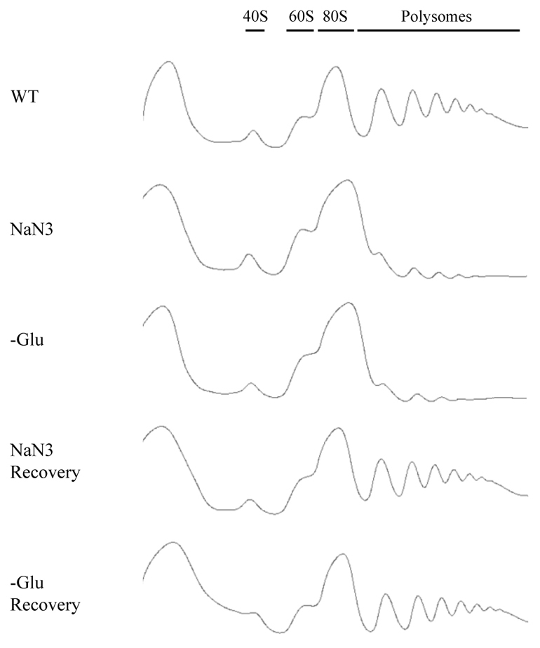
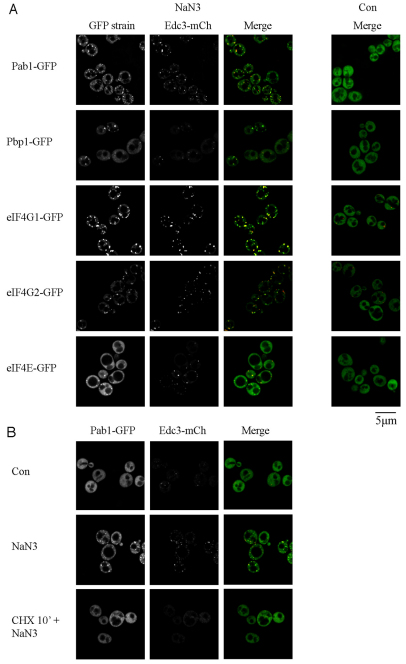
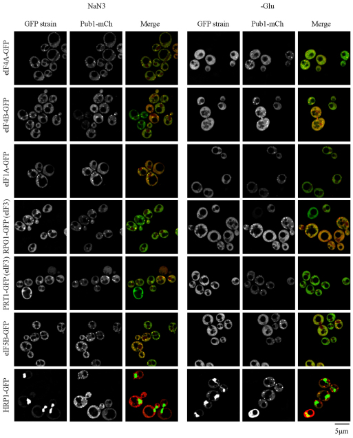
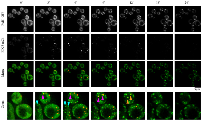
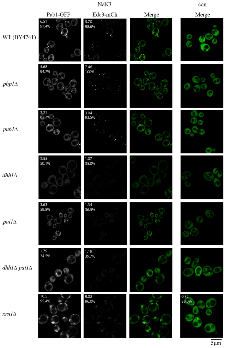
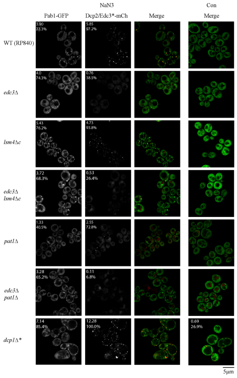
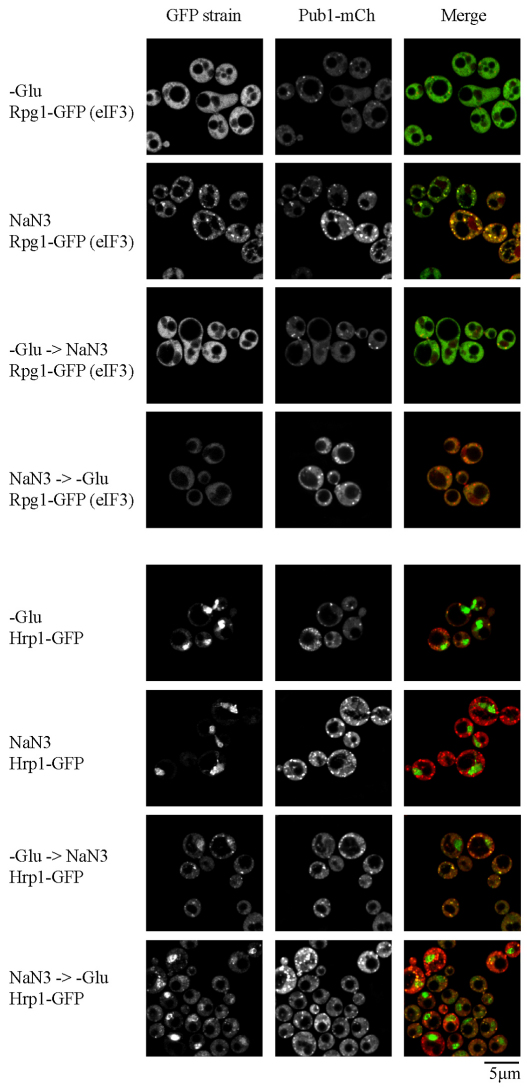
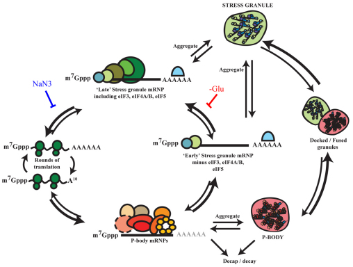
Eukaryotic cells respond to cellular stresses by the inhibition of translation and the accumulation of mRNAs in cytoplasmic RNA-protein (ribonucleoprotein) granules termed stress granules and P-bodies. An unresolved issue is how different stresses affect formation of messenger RNP (mRNP) granules. In the present study, we examine how sodium azide (NaN(3)), which inhibits mitochondrial respiration, affects formation of mRNP granules as compared with glucose deprivation in budding yeast. We observed that NaN(3) treatment inhibits translation and triggers formation of P-bodies and stress granules. The composition of stress granules induced by NaN(3) differs from that of glucose-deprived cells by containing eukaryotic initiation factor (eIF)3, eIF4A/B, eIF5B and eIF1A proteins, and by lacking the heterogeneous nuclear RNP (hnRNP) protein Hrp1. Moreover, in contrast with glucose-deprived stress granules, NaN(3)-triggered stress granules show different assembly rules, form faster and independently from P-bodies and dock or merge with P-bodies over time. Strikingly, addition of NaN(3) and glucose deprivation in combination, regardless of the order, always results in stress granules of a glucose deprivation nature, suggesting that both granules share an mRNP remodeling pathway. These results indicate that stress granule assembly, kinetics and composition in yeast can vary in a stress-specific manner, which we suggest reflects different rate-limiting steps in a common mRNP remodeling pathway.


Figures

Fig. 1.
Fig. 1.
NaN3 stress represses translation. Exponential-phase BY4741 cells were subject to control (WT) or stress conditions for 10 minutes [NaN3 at 0.5% (v/v) or glucose deprivation, –Glu], and stress conditions followed by recovery for 10 minutes in fresh medium. Global translation was monitored by polysome analysis.
Fig. 2.
Fig. 2.
NaN3 induces stress granules and P-bodies that require non-translating mRNA for assembly. (A) Exponential-phase cells expressing chromosomal GFP-tagged proteins and Edc3–mCh (pRP1574) were subjected to 0.5% (v/v) NaN3 for 30 minutes and examined. No GFP foci and only faint P-bodies were observed in glucose-containing control (Con) conditions, hence display of the merge image only. (B) Exponential-phase BY4741 cells expressing pRP1657 were subject to control or NaN3 treatment as above, or treatment with 100 μg/ml cycloheximide (CHX) 10 minutes before NaN3 treatment.
Fig. 3.
Fig. 3.
NaN3 stress granules harbor additional components associated with later stages of translation initiation. Exponential-phase cells expressing chromosomal GFP-tagged proteins and Pub1–mCh (pRP1661) were subjected to 0.5% (v/v) NaN3 for 30 minutes, or glucose deprivation (–Glu) stress, and examined. Under control conditions (not shown), none of the GFP-tagged factors formed cytoplasmic foci, whereas only faint P-bodies were observed.
Fig. 4.
Fig. 4.
Kinetic analysis of NaN3 stress granule and P-body formation. Exponential-phase BY4741, transformed with pRP1657, were subjected to NaN3 stress and immediately spotted onto microscopy slides for examination. Still images are taken from supplementary material Movie 1. Turquoise arrowheads indicate stress granule foci initially forming separately from visible P-bodies, whereas magenta arrowheads indicate P-body-associated stress granule formation. These stress granules often remain docked before, in some cases, fusing more completely with P-bodies (orange arrowheads). Note that, at later timepoints (at 18 and 24 minutes), significant photobleaching of the P-body signal has occurred, giving a false impression of P-body disappearance.
Fig. 5.
Fig. 5.
Null strain analysis of factors affecting NaN3 stress granule assembly. Exponential-phase cells, all of the BY4741 strain background, were transformed with plasmid pRP1657 and subjected to NaN3 stress [0.5% (v/v) for 30 minutes] or control conditions. Stress granules (Pab1–GFP) and P-body numbers (Edc3–mCh) were quantified in a blind manner. The number given in the top-left corner indicates the mean number of foci per cell, the percentage indicates the proportion of cells with one or more foci (see also supplementary material Tables S1, S2). con, control.
Fig. 6.
Fig. 6.
Null strain analysis of factors affecting NaN3 stress granule assembly. Exponential-phase cells of yRP840 background were transformed with pRP1660, except for the dcp1Δ strain, which was transformed with pRP1657. NaN3 stress and quantification were conducted as in Fig. 5. Con, control.
Fig. 7.
Fig. 7.
Combinatorial stress analysis reveals the dominance of glucose deprivation stress over NaN3 stress with regard to stress granule composition. Exponential-phase Rpg1–GFP or Hrp1–GFP cells, transformed with plasmid pRP1661, were subjected to each stress condition individually or in combination (see the Materials and Methods section). –Glu, glucose deprivation.
Fig. 8.
Fig. 8.
Theoretical models of mRNA remodeling during glucose deprivation and NaN3 stress. Glucose deprivation (–Glu) and NaN3 stress granules could in principle form through distinct assembly pathways, which are dependent and independent, respectively, of visible P-body aggregation. Alternatively, different rate-limiting steps in a cycle of broadly conserved mRNP remodeling events could lead to the formation of subtly different, but related, granules (e.g. ‘early’ and ‘late’ stress granules) during glucose deprivation and NaN3 stress, as indicated by the proposed points at which glucose deprivation and NaN3 stress could block the mRNA cycle on the basis of our observations. Note, poly(A) tails are shown in light gray within P-bodies to highlight the fact that P-body mRNPs are likely to be a mixture of adenylated and deadenylated species, the latter of which are more likely to undergo decay.

References

    1. Anderson P., Kedersha N. (2009). RNA granules: post-transcriptional and epigenetic modulators of gene expression. Nat. Rev. Mol. Cell Biol. 10, 430-436 - PubMed
    1. Brengues M., Parker R. (2007). Accumulation of polyadenylated mRNA, Pab1p, eIF4E, and eIF4G with P-bodies in Saccharomyces cerevisiae. Mol. Biol. Cell 18, 2592-2602 - PMC - PubMed
    1. Brengues M., Teixeira D., Parker R. (2005). Movement of eukaryotic mRNAs between polysomes and cytoplasmic processing bodies. Science 310, 486-489 - PMC - PubMed
    1. Buchan J. R., Parker R. (2009). Eukaryotic stress granules: the ins and outs of translation. Mol. Cell 36, 932-941 - PMC - PubMed
    1. Buchan J. R., Muhlrad D., Parker R. (2008). P bodies promote stress granule assembly in Saccharomyces cerevisiae. J. Cell Biol. 183, 441-455 - PMC - PubMed

Publication types

MeSH terms

LinkOut - more resources