

RCD1 and SRO1 are necessary to maintain meristematic fate in Arabidopsis thaliana
- PMID: 21172813
- PMCID: PMC3022410
- DOI: 10.1093/jxb/erq363
RCD1 and SRO1 are necessary to maintain meristematic fate in Arabidopsis thaliana
Abstract
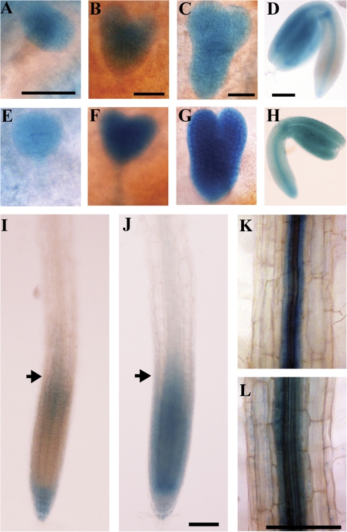
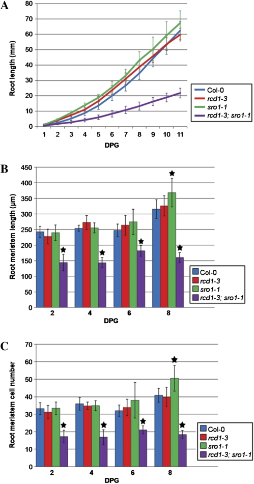
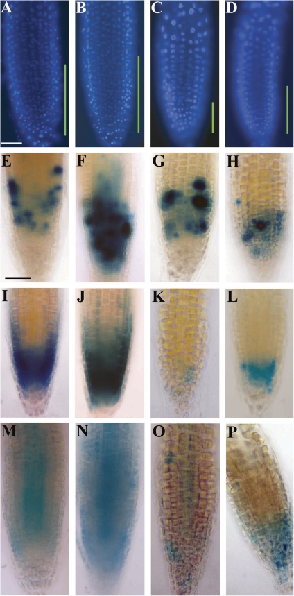
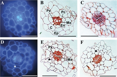
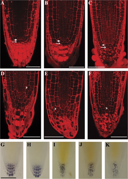
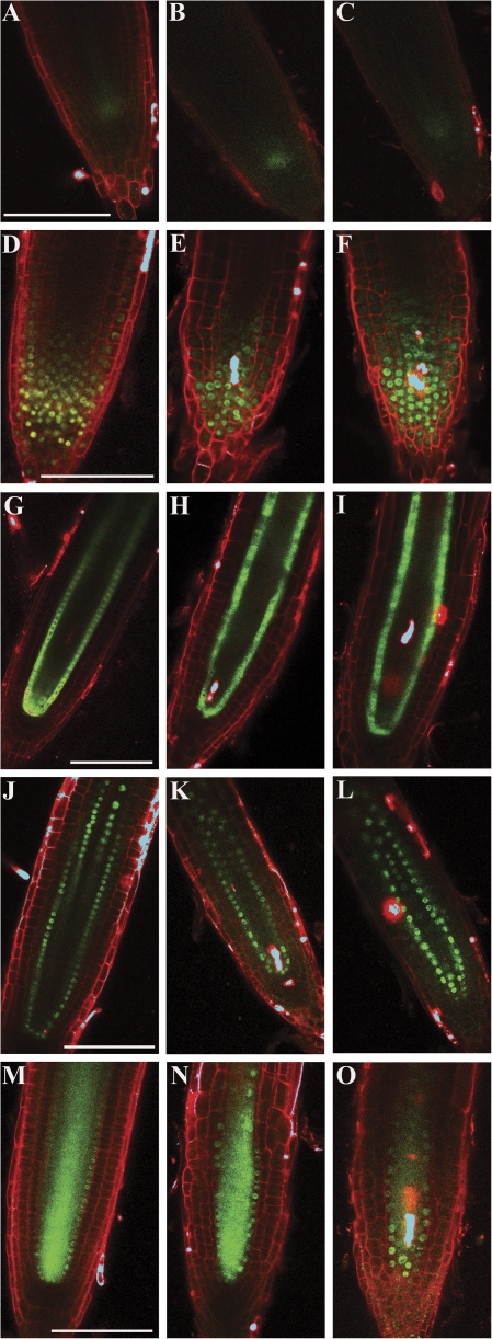
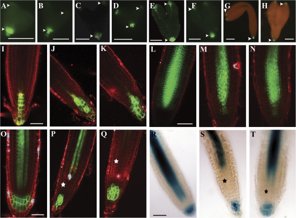
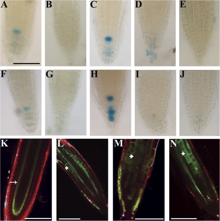
The radical-induced cell death1 and similar to RCD ONE1 genes of Arabidopsis thaliana encode members of the poly(ADP-ribose) polymerase (PARP) superfamily and have pleiotropic functions in development and abiotic stress response. In order to begin to understand the developmental and molecular bases of the defects seen in rcd1-3; sro1-1 plants, this study used the root as a model. Double mutant roots are short and display abnormally organized root apical meristems. However, acquisition of most cell fates within the root is not significantly disrupted. The identity of the quiescent centre is compromised, the zone of cell division is smaller than in wild-type roots and abnormal divisions are common, suggesting that RCD1 and SRO1 are necessary to maintain cells in a division-competent state and to regulate division plane placement. In addition, differentiation of several cell types is disrupted in rcd1-3; sro1-1 roots and shoots, demonstrating that RCD1 and SRO1 are also necessary for proper cell differentiation. Based on the data shown in this article and previous work, we hypothesize that RCD1 and SRO1 are involved in redox control and, in their absence, an altered redox balance leads to abnormal development.
Figures
Similar articles
-
The paralogous genes RADICAL-INDUCED CELL DEATH1 and SIMILAR TO RCD ONE1 have partially redundant functions during Arabidopsis development.Plant Physiol. 2009 Sep;151(1):180-98. doi: 10.1104/pp.109.142786. Epub 2009 Jul 22. Plant Physiol. 2009. PMID: 19625634 Free PMC article.
-
Unequally redundant RCD1 and SRO1 mediate stress and developmental responses and interact with transcription factors.Plant J. 2009 Oct;60(2):268-79. doi: 10.1111/j.1365-313X.2009.03951.x. Epub 2009 Jun 22. Plant J. 2009. PMID: 19548978
-
TPR5 is involved in directional cell division and is essential for the maintenance of meristem cell organization in Arabidopsis thaliana.J Exp Bot. 2016 Apr;67(8):2401-11. doi: 10.1093/jxb/erw043. Epub 2016 Feb 17. J Exp Bot. 2016. PMID: 26889009 Free PMC article.
-
Network building: transcriptional circuits in the root.Curr Opin Plant Biol. 2004 Oct;7(5):582-8. doi: 10.1016/j.pbi.2004.07.010. Curr Opin Plant Biol. 2004. PMID: 15337102 Review.
-
The gene regulatory network for root epidermal cell-type pattern formation in Arabidopsis.J Exp Bot. 2009;60(5):1515-21. doi: 10.1093/jxb/ern339. Epub 2009 Jan 27. J Exp Bot. 2009. PMID: 19174459 Free PMC article. Review.
Cited by
-
Metabolite Profiling of Paraquat Tolerant Arabidopsis thaliana Radical-induced Cell Death1 (rcd1)-A Mediator of Antioxidant Defence Mechanisms.Antioxidants (Basel). 2022 Oct 15;11(10):2034. doi: 10.3390/antiox11102034. Antioxidants (Basel). 2022. PMID: 36290757 Free PMC article.
-
Wild emmer introgression alters root-to-shoot growth dynamics in durum wheat in response to water stress.Plant Physiol. 2021 Nov 3;187(3):1149-1162. doi: 10.1093/plphys/kiab292. Plant Physiol. 2021. PMID: 34618034 Free PMC article.
-
The membrane-bound NAC transcription factor ANAC013 functions in mitochondrial retrograde regulation of the oxidative stress response in Arabidopsis.Plant Cell. 2013 Sep;25(9):3472-90. doi: 10.1105/tpc.113.117168. Epub 2013 Sep 17. Plant Cell. 2013. PMID: 24045019 Free PMC article.
-
Functions of the poly(ADP-ribose) polymerase superfamily in plants.Cell Mol Life Sci. 2012 Jan;69(2):175-89. doi: 10.1007/s00018-011-0793-4. Epub 2011 Aug 23. Cell Mol Life Sci. 2012. PMID: 21861184 Free PMC article. Review.
-
Spatially expressed WIP genes control Arabidopsis embryonic root development.Nat Plants. 2022 Jun;8(6):635-645. doi: 10.1038/s41477-022-01172-4. Epub 2022 Jun 16. Nat Plants. 2022. PMID: 35710883
References
-
- Adams-Phillips L, Wan J, Tan X, Dunning FM, Meyers BC, Michelmore RW, Bent AF. Discovery of ADP-ribosylation and other plant defense pathway elements through expression profiling of four different Arabidopsis-Pseudomonas R-avr interactions. Molecular Plant–Microbe Interactions. 2008;21:646–657. - PubMed
-
- Ahlfors R, Brosché M, Kollist H, Kangasjärvi J. Nitric oxide modulates ozone-induced cell death, hormone biosynthesis and gene expression in Arabidopsis thaliana. The Plant Journal. 2008 Nov 28 [Epub ahead of print] - PubMed
-
- Aida M, Beis D, Heidstra R, Willemsen V, Blilou I, Galinha C, Nussaume L, Noh YS, Amasino R, Scheres B. The PLETHORA genes mediate patterning of the Arabidopsis root stem cell niche. Cell. 2004;119:109–120. - PubMed
MeSH terms
Substances
LinkOut - more resources
Full Text Sources
Other Literature Sources
Molecular Biology Databases
