. 2011 Jan 1;124(Pt 1):133-43.
doi: 10.1242/jcs.072686.

An endophilin-dynamin complex promotes budding of clathrin-coated vesicles during synaptic vesicle recycling

Affiliations

An endophilin-dynamin complex promotes budding of clathrin-coated vesicles during synaptic vesicle recycling

Anna Sundborger et al. J Cell Sci. .

Abstract

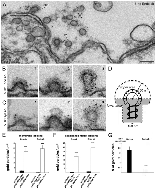
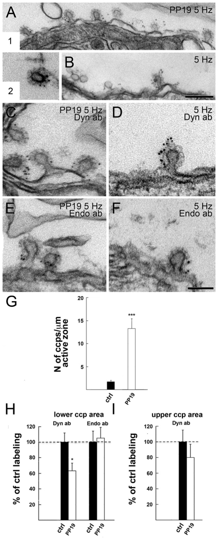
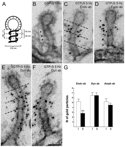
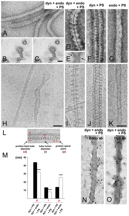
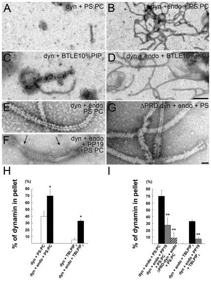
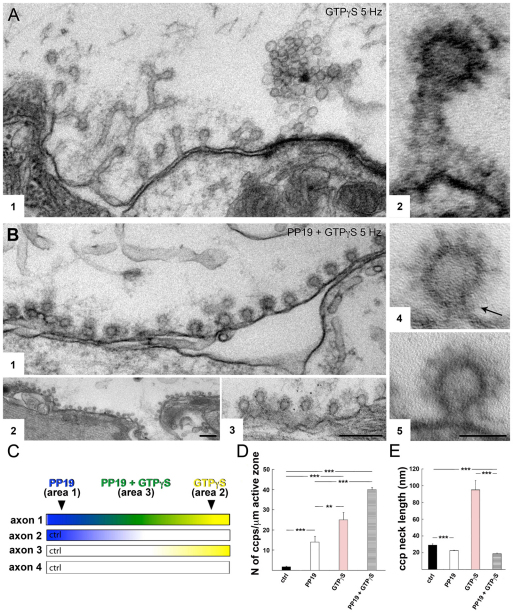
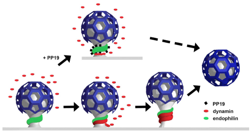
Clathrin-mediated vesicle recycling in synapses is maintained by a unique set of endocytic proteins and interactions. We show that endophilin localizes in the vesicle pool at rest and in spirals at the necks of clathrin-coated pits (CCPs) during activity in lamprey synapses. Endophilin and dynamin colocalize at the base of the clathrin coat. Protein spirals composed of these proteins on lipid tubes in vitro have a pitch similar to the one observed at necks of CCPs in living synapses, and lipid tubules are thinner than those formed by dynamin alone. Tubulation efficiency and the amount of dynamin recruited to lipid tubes are dramatically increased in the presence of endophilin. Blocking the interactions of the endophilin SH3 domain in situ reduces dynamin accumulation at the neck and prevents the formation of elongated necks observed in the presence of GTPγS. Therefore, endophilin recruits dynamin to a restricted part of the CCP neck, forming a complex, which promotes budding of new synaptic vesicles.


Figures

Fig. 1.
Fig. 1.
Endophilin accumulates at the periactive zone after stimulation and colocalizes with dynamin at the necks of CCPs. (A) TEM image of the periactive zone of a reticulospinal synapse in lamprey stimulated at 5 Hz for 20 minutes and labeled with antibodies against endophilin. Gold particles are accumulated at CCPs and vesicles (sv) in the periactive zone. (B,C) Clathrin-coated intermediates labeled with antibodies against endophilin (B) and dynamin (C) at higher magnification (1 – shallow, 2 – non-constricted, and 3 – constricted states). (D) Schematic illustration of a constricted CCP divided into upper and lower areas used for the quantifications of gold particle labeling. (E) Bar graph showing the densities of gold particle labeling for endophilin and dynamin in the region 15 nm from the presynaptic membrane in the periactive zone (500 nm from the active zone) and outside synaptic regions. (F) Bar graph showing gold particle labeling for endophilin and dynamin in the axoplasmic matrix 500 μm2 lateral to the active zone and outside the synaptic region. (G) Bar graph showing the ratios of gold particle densities in the ‘upper’ versus ‘lower’ areas of the constricted CCPs labeled for endophilin and dynamin. (D,G) Quantification of immunogold labeling on constricted CCPs shows that endophilin predominantly resides at the lower half of CCPs (upper:lower ratio=0.37±0.17), whereas dynamin is found more frequently in the upper half of constricted CCPs (upper:lower ratio=2.3±0.35). Bars represent means ± s.e.m. (***P<0.001, Student's t-test; n=35 CCPs). Scale bars: 100 nm (A); 50 nm (B,C).
Fig. 2.
Fig. 2.
Acute perturbation of endophilin SH3-domain interactions perturbs localization of dynamin at necks of CCPs and inhibits fission in living synapses. (A1) Electron micrograph showing a periactive zone of a lamprey synapse stimulated at 5 Hz for 20 minutes after microinjection of PP19 and labeling with antibodies against dynamin. (A2) Free clathrin-coated vesicle found in the injected synapse shown in A1, but here at high magnification. (B) TEM image of a periactive zone in a control, non-injected synapse stimulated at 5 Hz and labeled with antibodies against dynamin. (C–F) TEM images of CCPs from stimulated synapses in axons microinjected with PP19 and labeled with antibodies against dynamin (C) and endophilin (E) and in stimulated control axons labeled with the same antibodies (D and F, respectively). (G) Bar graph showing an accumulation of CCPs in synapses injected with PP19 as compared with control synapses (P<0.001, Student's t-test; n=6–8 synapses). (H) Bar graph showing dynamin and endophilin labeling in the lower area of CCPs in synapses injected with PP19 and stimulated at 5 Hz for 20 minutes compared with that of stimulated control synapses from the same preparation. (I) Bar graph showing dynamin labeling in the upper area of CCPs in synapses injected with PP19 and stimulated at 5 Hz for 20 minutes compared with that of stimulated control synapses from the same preparation (*P<0.05, Student's t-test; n=35 CCPs). Bars in (G,H,I) represent means ± s.e.m. Scale bars: 50 nm (A2,C–F); 200 nm (A1,B).
Fig. 3.
Fig. 3.
Endophilin and dynamin colocalize in the region proximal to the coat of elongated constricted necks induced by GTPγS. (A) Schematic illustration of areas 1 (‘I’) and 2 (‘II’) of the CCP neck defined for quantifications of antibody labeling. (B) TEM image of a constricted CCP in the periactive zone of a synapse injected with GTPγS and stimulated at 5 Hz for 20 minutes. Note the presence of dense rings (arrowheads). (C,D) and (E,F) Electron micrographs of the CCPs labeled with antibodies against endophilin and dynamin, respectively. Areas ‘I’ and ‘II’ are marked by dashed lines. (G) Bar graph showing the number of gold particles labeling endophilin, dynamin and amphiphysin in the two regions defined in (A). Bars represent means ± s.e.m. (***P<0.001, Student's t-test; n=35 CCPs). Scale bar: 50 nm (B–F).
Fig. 4.
Fig. 4.
Ultrastructural characterization of the endophilin–dynamin complex in vitro. (A,H) TEM images of PS liposomes tubulated by endophilin and dynamin and visualized by negative staining (A) and cryo-EM (H). (B,C) Stereo pair of TEM images of CCPs taken at ± 4° tilts from synapses injected with GTPγS and stimulated at 5 Hz. (D,I) High magnifications of endophilin–dynamin-decorated PS tubes in negative stain (D) and vitreous ice (I). Note the regular arrangement of the protein complex and its structural similarity to the protein complex assembled on the neck of a constricted CCP (E, arrows). (F,J) TEM images of dynamin-decorated PS tubes in negative stain (F) and vitreous ice (J). Note that the diameter of the protein–lipid tube in F and J is larger than the one shown in D and I. (G,K) TEM images of tubulated PS liposomes decorated by endophilin visualized by negative stain (G) and cryo-EM (K). (L) Measurements of protein tube diameter (d), tube lumen diameter (l) and protein spiral pitch (p), measured in protein–lipid tubes embedded in amorphous ice quantified in the bar graph shown in (M). Note the statistically significant difference in each case between dynamin-decorated tubes and tubes decorated with dynamin and endophilin (***P<0.001, Student's t-test; n=100 tubes). (N,O) Electron micrographs of PS liposomes decorated by endophilin–dynamin spirals and labeled with antibodies against endophilin and dynamin, respectively. Scale bars: 100 nm (A,H); 50 nm (B,C); 50 nm (D–G,I–K,N,O).
Fig. 5.
Fig. 5.
Endophilin–dynamin complex formation facilitates recruitment of dynamin to liposomes. (A,B) TEM images of PS:PC liposomes decorated by dynamin alone (A) or decorated with endophilin and dynamin in negative stain (B). (C,D) TEM images of total-brain-lipid plus 10% PIP2 liposomes (BTLEPIP2) decorated with dynamin (C) or endophilin–dynamin complexes (D). Note an increase in the number of tubules in (B) and (D) compared with A, and C. (E,F) Electron micrographs of PS:PC liposomes in the presence of endophilin and dynamin (E) and following addition of 300 μM PP19 (F). Note the gaps in the decoration exposing naked liposomes (arrows). (G) TEM image of PS liposomes in the presence of endophilin and ΔPRD-dynamin. Note the absence of well-ordered complexes seen in (E). (H) Bar graph shows the increase in the amount of dynamin (expressed as a percentage) in the pellet fraction after mixing with PS:PC or BTLEPIP2 in the absence (white bars) and presence of endophilin (black bars). (I) Bar graph shows reduction in the amount of dynamin (expressed as a percentage) in the pellet fraction after mixing with PS:PC or BTLEPIP2 and endophilin (black bars) in the absence and presence of PP19 (grey bars) and following deletion of its PRD (hatched bar). Bars show means ± s.e.m. (*P<0.05, **P<0.01, Student's t-test; n=8 experiments for PS:PC and 2 for BTLEPIP2). Scale bars: 500 nm (A–D), 50 nm (E–G).
Fig. 6.
Fig. 6.
PP19 prevents the formation of elongated necks decorated with dynamin-containing spirals induced by GTPγS. (A) Control TEM image of the periactive zone of a reticulospinal synapse from an axon microinjected with GTPγS and Texas Red (see panel C, ‘axon 3’, ‘area 3’). Coated pits with elongated necks decorated by spirals are seen accumulated in the periactive zone. (A2) High-magnification image of a constricted coated pit with an elongated neck from the synapse shown in A1. Note the thick spiral. (B) Electron micrograph of the periactive zone of a reticulospinal synapse injected with both PP19 and GTPγS (see panel C, ‘axon 1’, ‘area 3’). Note the accumulation of CCPs with short necks in the periactive zone (B1,2). (B3) TEM image illustrating dynamin immunogold labeling of constricted CCPs with short necks in a synapse injected with PP19 and GTPγS. (B4,5) High-magnification images of constricted coated pits with short necks from the synapse shown in (B1) (arrow indicates a thin spiral). (C) Schematics of the microinjection experiment. The zone ‘area 1’ (blue) delineates the PP19 microinjection site, and ‘area 2’ (yellow) delineates the GTPγS microinjection site. The region ‘Area 3’ (green) marks where the two compounds mixed in the axon that was microinjected with both compounds. Control axons were injected with either PP19 (‘axon 2’) or GTPγS (‘axon 3’). (D) Bar graph showing the number of CCPs in the periactive zones of synapses microinjected with PP19, GTPγS, PP19 plus GTPγS or non-injected control axons stimulated at 5 Hz. Values are normalized to the length of the active zone. (E) Bar graph showing the length of necks of CCPs from the synapses described in (D). Statistical significance of differences between means ± s.e.m. was determined by a Student's t-test (**P<0.01, ***P<0.001; n=35 CCPs). Scale bars: 200 nm (A1,B1,B3), 50 nm (A2,B4,B5), 500 nm (B2).
Fig. 7.
Fig. 7.
Schematic illustrating the proposed function of the endophilin–dynamin interaction at the neck of CCPs during synaptic vesicle recycling. Dynamin and endophilin are recruited to different regions of the coated pit. Endophilin induces curvature of the neck and serves as a template for dynamin. An endophilin–dynamin fission complex forms at the base of the coat. This complex promotes recruitment of dynamin to the neck, which results in GTP-mediated fission. The presence of PP19 blocks the dynamin binding sites on endophilin, preventing formation of the complex and efficient recruitment of dynamin, resulting in perturbation of fission (dashed arrow).

References

    1. Andersson F., Löw P., Brodin L. (2010). Selective perturbation of the BAR domain of endophilin impairs synaptic vesicle endocytosis. Synapse 64, 556-560 - PubMed
    1. Anggono V., Robinson P. J. (2007). Syndapin I and endophilin I bind overlapping proline-rich regions of dynamin I: role in synaptic vesicle endocytosis. J. Neurochem. 102, 931-943 - PubMed
    1. Conibear E. (2010). Converging views of endocytosis in yeast and mammals. Curr. Opin. Cell Biol. 22, 513-518 - PubMed
    1. Cremona O., Di Paolo G., Wenk M. R., Luthi A., Kim W. T., Takei K., Daniell L., Nemoto Y., Shears S. B., Flavell R. A., et al. (1999). Essential role of phosphoinositide metabolism in synaptic vesicle recycling. Cell 99, 179-188 - PubMed
    1. Dickman D. K., Horne J. A., Meinertzhagen I. A., Schwarz T. L. (2005). A slowed classical pathway rather than kiss-and-run mediates endocytosis at synapses lacking synaptojanin and endophilin. Cell 123, 521-533 - PubMed

Publication types

MeSH terms