. 2011 May;28(5):1645-59.
doi: 10.1093/molbev/msq345. Epub 2010 Dec 20.

Novel protein genes in animal mtDNA: a new sex determination system in freshwater mussels (Bivalvia: Unionoida)?

Affiliations

Novel protein genes in animal mtDNA: a new sex determination system in freshwater mussels (Bivalvia: Unionoida)?

Sophie Breton et al. Mol Biol Evol. 2011 May.

Abstract

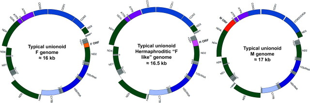
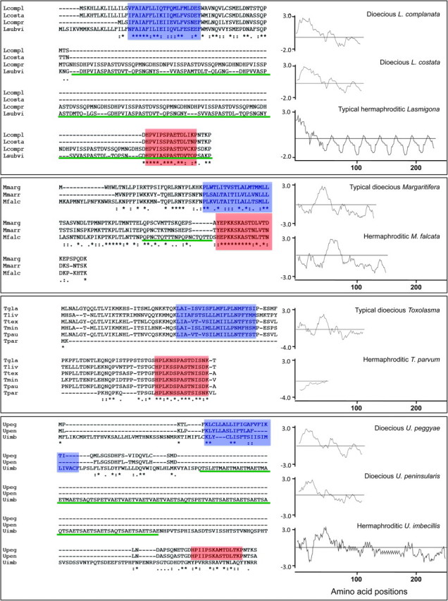
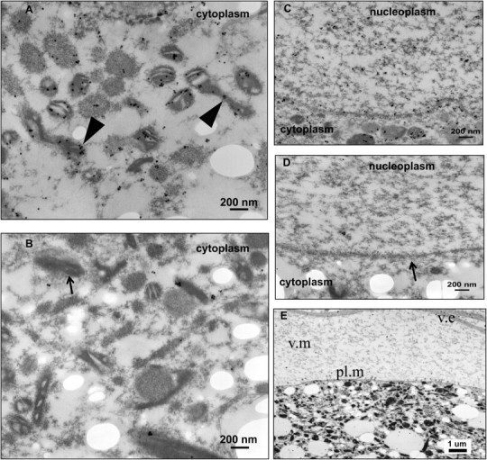
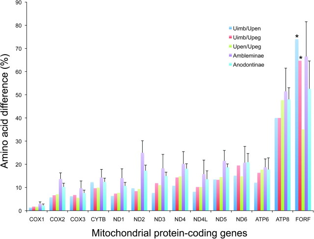
Mitochondrial (mt) function depends critically on optimal interactions between components encoded by mt and nuclear DNAs. mitochondrial DNA (mtDNA) inheritance (SMI) is thought to have evolved in animal species to maintain mito-nuclear complementarity by preventing the spread of selfish mt elements thus typically rendering mtDNA heteroplasmy evolutionarily ephemeral. Here, we show that mtDNA intraorganismal heteroplasmy can have deterministic underpinnings and persist for hundreds of millions of years. We demonstrate that the only exception to SMI in the animal kingdom, that is, the doubly uniparental mtDNA inheritance system in bivalves, with its three-way interactions among egg mt-, sperm mt- and nucleus-encoded gene products, is tightly associated with the maintenance of separate male and female sexes (dioecy) in freshwater mussels. Specifically, this mother-through-daughter and father-through-son mtDNA inheritance system, containing highly differentiated mt genomes, is found in all dioecious freshwater mussel species. Conversely, all hermaphroditic species lack the paternally transmitted mtDNA (=possess SMI) and have heterogeneous macromutations in the recently discovered, novel protein-coding gene (F-orf) in their maternally transmitted mt genomes. Using immunoelectron microscopy, we have localized the F-open reading frame (ORF) protein, likely involved in specifying separate sexes, in mitochondria and in the nucleus. Our results support the hypothesis that proteins coded by the highly divergent maternally and paternally transmitted mt genomes could be directly involved in sex determination in freshwater mussels. Concomitantly, our study demonstrates novel features for animal mt genomes: the existence of additional, lineage-specific, mtDNA-encoded proteins with functional significance and the involvement of mtDNA-encoded proteins in extra-mt functions. Our results open new avenues for the identification, characterization, and functional analyses of ORFs in the intergenic regions, previously defined as "noncoding," found in a large proportion of animal mt genomes.


Figures

F<sc>IG</sc>. 1.
FIG. 1.
Gene maps of the sex-associated and hermaphroditic “F-like” mt genomes of freshwater mussels. Gene identities: complex I in green; complex III in light blue; complex IV in blue; complex V in light purple; small and large ribosomal RNAs (in purple). Transfer RNAs (in gray) are depicted by one letter amino acid codes. F-ORF, F-specific open reading frame (orange); H-ORF, Hermaphroditic-specific open reading frame (pink); M-ORF, M-specific open reading frame (red). Genes positioned inside the circle are encoded on the light strand, and genes outside the circle are encoded on the heavy strand.
F<sc>IG</sc>. 2.
FIG. 2.
Primary and secondary structures of F-ORF and H-ORF amino acid sequences in dioecious and hermaphroditic freshwater mussel species, respectively. From above to below: Comparisons of dioecious Lasmigona spp. and hermaphroditic L. compressa and L. subviridis F- and H-ORFs. (Lcompl = L. complanata; Lcosta = L. costata; Lcompr = L. compressa; Lsubvi = L. subviridis). F-ORFs and H-ORF for Margaritifera margaritifera, M. marrianae and the hermaphroditic M. falcata. (Mmarg = M. margaritifera; Mmarr = M. marrianae; Mfalc = M. falcata). Comparisons of dioecious Toxolasma spp. and hermaphroditic T. parvum F-ORFs and H-ORF, respectively. (Tgla = T. glans; Tliv = T. lividus; Ttex = T. texasensis; Tmin = T. minor; Tpau = T. paulus; Tpar = T. parvum). F-ORFs and H-ORF for dioecious Utterbackia peggyae, U. peninsularis and hermaphroditic U. imbecillis. (Upeg = U. peggyae; Upen = U. peninsularis; Uimb = U. imbecillis). Alignment of amino acid sequences and secondary structures for the F- and H-ORFs are shown on the left. Color highlighting is as follows: blue box, the predicted transmembrane domain (TMH); reddish orange box, the relatively conserved, near-C-terminus, 15 amino acid motif; green, repetitive units. Hydropathy profiles are shown on the right. Numbers below profiles designate amino acid positions in each protein.
F<sc>IG</sc>. 3.
FIG. 3.
BI MAP tree from analysis of concatenated Fcox1/nd1 DNA sequences using Hyridella menziesi (Unionoida: Hyriidae) as the outgroup. An asterisk above a branch = BI posterior probability (×100) ≥ 95. An asterisk below a branch = ML BSP (1000×) ≥ 70. Black indicates lineages that are dioecious, contain an F-orf in the F mt genome and display DUI (=M mt genome present). Orange indicates lineages that are hermaphroditic, contain an H-orf in the F mt genome and display SMI (=M mt genome absent). Hermaphroditism, H-orfs and SMI concurrently evolved four distinct times. Proportional likelihood values (all nodes significant) are given in rectangles for the character state estimates at eight nodes (orange diamonds, character states = hermaphroditism, H-orf and M genome absent; black diamonds, character states = dioecy, F-orf, and M genome present, BSP = Bootstrap Support Percentages).
F<sc>IG</sc>. 4.
FIG. 4.
Immunoelectron microscopy (IEM) shows the presence of the F-ORF protein in the eggs of the dioecious species V. ellipsiformis. (A and C) Female-transmitted F-ORF is localized to the cytoplasm (mitochondria). (C) Female-transmitted F-ORF is localized to the nuclear membrane and nucleoplasm. (B and D) negative control IEM photomicrographs. (Note: black arrowheads in figure 4A indicate the presence of the F-ORF protein in mitochondria, small black arrow in figure 4B indicates a mitochondrion, small black arrow in figure 4D indicates the nuclear membrane). (E) Section of an egg showing egg plasma membrane (pl.m), vitelline matrix (v.m), and vitelline envelope (v.e).
F<sc>IG</sc>. 5.
FIG. 5.
The extent of amino acid divergence (% amino acid difference) for each mt protein coding gene between U. imbecillis and U. peninsularis (Uimb/Upen), U. imbecillis and U. peggyae (Uimb/Upeg), U. peninsularis and U. peggyae (Upen/Upeg), and among species of the subfamilies Ambleminae (i.e., H. cumingii, L. ornata, Q. quadrula, T. parvum, V. ellipsiformis), and Anodontinae (i.e., C. plicata, L. compressa, Pygadodon grandis, U. imbecillis, U. peninsularis, U. peggyae).*F-ORF/H-ORF comparisons. (H-ORF protein sequences have been excluded from the analyses of Ambleminae and Anodontinae).

References

    1. Altschul SF, Madden TL, Schaffer AA, Zhang J, Zhang Z, Miller W, Lipman DJ. Gapped BLAST and PSI-BLAST: a new generation of protein database search programs. Nucleic Acids Res. 1997;25:3389–3402. - PMC - PubMed
    1. Arai M, Mitsuke H, Ikeda M, Xia JX, Kikuchi T, Satake M, Shimizu T. ConPred II: a consensus prediction method for obtaining transmembrane topology models with high reliability. Nucleic Acids Res. 2004;32:W390–W393. - PMC - PubMed
    1. Ballard JWO, Whitlock MC. The incomplete natural history of mitochondria. Mol Ecol. 2004;13:729–744. - PubMed
    1. Barker D, Pagel M. Predicting functional gene links from phylogenetic-statistical analyses of whole genome. PLoS Comput Biol. 2005;1:e3. - PMC - PubMed
    1. Benson D, Karsch-Mizrachi I, Lipman D, Ostell J, Wheeler D. GenBank update. Nucleic Acids Res. 2004;32:D23–D26. - PMC - PubMed

Publication types

MeSH terms

Substances