. 2011 Feb;155(2):735-50.
doi: 10.1104/pp.110.168757. Epub 2010 Dec 20.

Arabidopsis homologs of the petunia hairy meristem gene are required for maintenance of shoot and root indeterminacy

Affiliations

Arabidopsis homologs of the petunia hairy meristem gene are required for maintenance of shoot and root indeterminacy

Eric M Engstrom et al. Plant Physiol. 2011 Feb.

Abstract

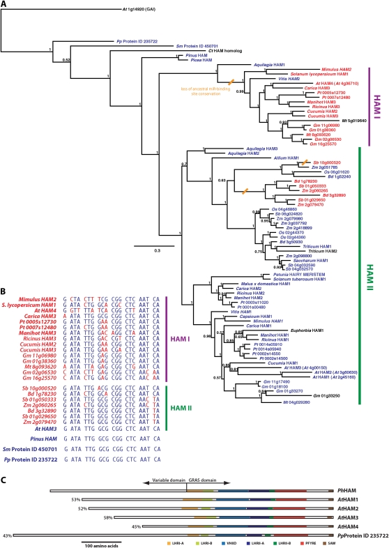
Maintenance of indeterminacy is fundamental to the generation of plant architecture and a central component of the plant life strategy. Indeterminacy in plants is a characteristic of shoot and root meristems, which must balance maintenance of indeterminacy with organogenesis. The Petunia hybrida HAIRY MERISTEM (HAM) gene, a member of the GRAS family of transcriptional regulators, promotes shoot indeterminacy by an undefined non-cell-autonomous signaling mechanism(s). Here, we report that Arabidopsis (Arabidopsis thaliana) mutants triply homozygous for knockout alleles in three Arabidopsis HAM orthologs (Atham1,2,3 mutants) exhibit loss of indeterminacy in both the shoot and root. In the shoot, the degree of penetrance of the loss-of-indeterminacy phenotype of Atham1,2,3 mutants varies among shoot systems, with arrest of the primary vegetative shoot meristem occurring rarely or never, secondary shoot meristems typically arresting prior to initiating organogenesis, and inflorescence and flower meristems exhibiting a phenotypic range extending from wild type (flowers) to meristem arrest preempting organogenesis (flowers and inflorescence). Atham1,2,3 mutants also exhibit aberrant shoot phyllotaxis, lateral organ abnormalities, and altered meristem morphology in functioning meristems of both rosette and inflorescence. Root meristems of Atham1,2,3 mutants are significantly smaller than in the wild type in both longitudinal and radial axes, a consequence of reduced rates of meristem cell division that culminate in root meristem arrest. Atham1,2,3 phenotypes are unlikely to reflect complete loss of HAM function, as a fourth, more distantly related Arabidopsis HAM homolog, AtHAM4, exhibits overlapping function with AtHAM1 and AtHAM2 in promoting shoot indeterminacy.


Figures

Figure 1.
Figure 1.
Arabidopsis HAM homolog proteins occupy two distantly related flowering plant HAM protein clades, characterized by retention or loss of microRNA regulation. A, Phylogeny of HAM proteins. The rooted phylogram shows aligned protein sequences of 78 HAM proteins from 23 flowering plant species, the gymnosperms Pinus and Picea, the fern Ceratopteris thalictroides, the lycophyte S. moellendorffii, and the moss P. patens (Supplemental Table S1), derived from Bayesian inference. Support values, indicated to the left of nodes, denote posterior probabilities. The scale bar indicates substitutions per site. The tree is rooted with a set of 12 DELLA proteins, although for visual simplicity only the Arabidopsis GA-INSENSITIVE protein is shown. Proteins encoded by genes that retain a perfectly conserved MIR-binding sequence are colored blue; proteins encoded by genes in which the MIR-binding sequence is imperfectly conserved are colored red. The phylogenetic locations of inferred losses in MIR-binding sequence conservation are indicated by slanted orange bars. The two largest monophyletic clades of flowering plant HAM proteins are designated HAM I and HAM II. B, Evolution of the MIR-binding sequence in flowering plant HAM genes. The MIR170/171-binding site sequence of AtHAM3 (Llave et al., 2002) is shown, along with the homologous sequences of HAM I and HAM II genes that deviate from the ancestral MIR-binding sequence and the conserved MIR-binding sequences of Pinus, Selaginella, and Physcomitrella. Nucleotides conserved with the ancestral MIR-binding sequence are colored blue; nucleotides that deviate from the ancestral MIR-binding sequence are colored red. C, Relative amino acid sequence identity of Arabidopsis homologs to Petunia HAM. A more distantly related HAM homolog from Physcomitrella is included for comparison. Percentage of pairwise amino acid identity between the aligned C-terminal GRAS domain with Petunia HAM, excluding alignment gaps, is indicated to the left of each Arabidopsis and Physcomitrella homolog. Indicated GRAS domain subunits follow the criteria proposed by Tian et al. (2004).
Figure 2.
Figure 2.
Arabidopsis HAM orthologs are expressed in meristematic and differentiated tissues of both the shoot and the root. A, In situ localization of AtHAM1 in a Ler shoot apex in a median longitudinal section. The L1 meristem cell layer is indicated with the arrowhead. A strong signal is consistently detected in lateral organ primordia. Signal is also detected in the meristem itself, with the highest level of signal present in the basal meristem regions and reduced or no signal in evidence in the uppermost cell layers of the central meristem region. AtHAM1 expression in the provasculature is indicated with the arrow. B, Expression maps of Arabidopsis HAM orthologs in root tissue, from The Arabidopsis Gene Expression Database (Birnbaum et al., 2003; Brady et al., 2007). Darker hues reflect higher relative expression levels within the root and between AtHAM orthologs. C, Relative expression levels of AtHAM orthologs in specific cell and tissue types. Values graphed are means of three replicates of normalized expression levels derived from mixed-model ANOVA analysis profiled by microarray profiling. Specific cell and tissue types are indicated, along with the marker employed to delineate spatial expression patterns in parentheses. Data shown are derived from the analysis reported by Brady et al. (2007), with the exceptions of cortex/endodermal initial (CEI; Sozzani et al., 2010) and mature endodermis (Mat ENDO; Carlsbecker et al., 2010). QC indicates the quiescent center.
Figure 3.
Figure 3.
Atham1,2,3 mutants exhibit arrest and differentiation of secondary meristems and altered structure and function of the primary shoot apical meristem. A, RT-PCR analysis of wild-type Wassilewskija (Ws) and a homozygous Atham1-1 mutant with primers flanking the T-DNA insertion site (Supplemental Table S2). Primers designed to amplify ACTIN2 cDNA were employed as a control for cDNA quality. B, RT-PCR analysis of wild-type Ler and homozygous Atham2-1 and Atham3-1 mutants with primers flanking the Ds insertion sites (Supplemental Table S2). Amplification from genomic DNA (gDNA) is employed as a reference for product size and primer set efficacy. AtHAM2 and AtHAM3 primer sets serve as reciprocal controls for cDNA quality in Ler, Atham2-1, and Atham3-1 genotypes. C, Ler and an Atham1,2,3 mutant at 3 d postgermination on sterile medium. Atham1,2,3 mutants exhibit elongation of the primary root, demonstrating the presence of a root meristem. The hypocotyl and cotyledons are evident in Atham1,2,3 mutants and do not differ significantly from the wild type in appearance, although epinastic curvature of the cotyledons is common in Atham1,2,3 mutants. D, Ler and an Atham1,2,3 mutant at 12 d postgermination. Postembryonic leaves are labeled (numbering of leaves 1 and 2 is arbitrary, as these two leaves arise roughly simultaneously). By emergence of the fourth leaf, deviations from wild-type phyllotaxis are apparent in many Atham1,2,3 mutants. E, Longitudinal section through shoot apices of Ler and an Atham1,2,3 mutant at 12 d postgermination. Atham1,2,3 mutants consistently exhibit broader and flatter primary shoot apical meristems relative to the wild type at 12 d postgermination, but significant differences in meristem size in Atham1,2,3 mutants relative to the wild type are not evident. F, Set of fully expanded rosette leaves of Ler and an Atham1,2,3 mutant. Leaf 8 of Ler is largely missing from this set. Atham1,2,3 rosette leaves typically exhibit reduced laminar expansion relative to the wild type. G, Epidermal surfaces of fully expanded rosette leaves of Ler and an Atham1,2,3 mutant imaged by scanning electron microscopy. Increases in average epidermal cell surface area relative to the wild type are evident on both the adaxial (Ad) and abaxial (Ab) leaf surfaces of Atham1,2,3 mutants. H, Ler and Atham1,2,3 mutant shoot phenotypes postflowering. Secondary shoots are typically in evidence emerging from axils of both the rosette and inflorescence at this stage in the wild type but are rarely observed in Atham1,2,3 mutants. I and J, Surface of an arrested rosette axillary meristem of an Atham1,2,3 mutant visualized by scanning electron microscopy. The main inflorescence stem (IS) is indicated for positional reference in I. The arrowhead in J indicates a fully differentiated stomata. K, Radially symmetrical multicellular structure, indicated with the arrow, emerging from an arrested axillary inflorescence meristem of an Atham1,2,3 mutant, visualized by digital optical microscopy.
Figure 4.
Figure 4.
Inflorescence phenotypes of Atham1,2,3 mutants. A, Time until flowering, assessed by petal emergence from the first flower, in Ler and Atham1,2,3 mutants. Error bars indicate 95% confidence interval of the mean. B, Inflorescence apices of an arrested Atham1,2,3 mutant and Ler, viewed with scanning electron microscopy. An arrested flower meristem (FM) is indicated to the left of the inflorescence meristem (IM). C, Progression in loss of indeterminacy in an Atham1,2,3 inflorescence viewed with scanning electron microscopy. Floral organs are labeled as follows: gynoecium (G), stamen (St), petal (P), and sepal (Se). Flowers are labeled according to sequence of initiation (F1–F5). D, Median longitudinal sections through Ler and Atham1,2,3 mutant inflorescence apices stained with toluidine blue and visualized by bright-field microscopy. Flower primordia are indicated (FP). Wild-type meristem cells stain densely, indicating high concentrations of cytoplasm. The arrow indicates a vacuolated cell in the Atham1,2,3 L1 meristem layer. Supernumerary meristem layers are evident in the Atham1,2,3 meristem. E, Abnormal phyllotaxis and organ transformation in an Atham1,2,3 mutant inflorescence. Three cauline leaves have arisen in close spatial and temporal proximity (compare with Ler in Fig. 3H). A flower has developed in the axillary meristem of the right-most cauline leaf at the position normally occupied by a secondary inflorescence shoot. Epinastic curling of cauline leaves is typical of Atham1,2,3 mutants. F, Cross sections through the cauline leaf lamina of Ler and an Atham1,2,3 mutant stained with toluidine blue and visualized by bright-field microscopy. Double-arrowed lines represent the approximate median thickness of adjacent cauline leaves. G, Vascular bundles of Ler and Atham1,2,3 cauline leaves. Xylem (Xy) is positioned adaxial to phloem (Ph) in Atham1,2,3 cauline leaves.
Figure 5.
Figure 5.
Root phenotypes of Atham1,2,3 mutants. A, Ler, Atham1,2,3, and shr2 plants grown for 12 d following germination on sterile medium. Atham1,2,3 mutants exhibit comparable reductions in primary root length relative to the wild type as shr2 mutants. B, Primary root growth rate of Ler and Atham1,2,3 mutants grown for 12 d following germination on sterile medium. Error bars indicate 2× SEM. WT, Wild type. C, Lugol-stained roots of Ler and an Atham1,2,3 mutant at 9 d following germination viewed by bright-field microscopy. Purple staining indicates starch granules in differentiated columella cells. The position of the quiescent center is indicated with the arrowheads. The reduction in primary root diameter in Atham1,2,3 mutants relative to the wild type is evident. D to H, Optical cross sections through primary root apices of Ler and Atham1,2,3 mutants visualized by confocal microscopy. D, F, G, and H show longitudinal cross sections through the root meristems of Ler and Atham1,2,3 mutants at 9 d following germination. The boundary between the root meristem and elongation zone is indicated in F, with the white arrowhead in the Atham1,2,3 panel, and is outside the frame of the Ler panel. E, Radial cross sections of Ler and Atham1,2,3 mutant meristematic zones at 9 d following germination. Epidermis (Ep) and cortex (C) are indicated. G, An Atham1,2,3 mutant at 10 d following germination shows loss of meristem indeterminacy. H, Longitudinal section through the root apex of an Atham1,2,3 mutant exhibiting root meristem bifurcation. I, Root apices of Columbia (Col) and Atham1,2,3 mutant roots expressing the pDR5::GUS auxin reporter construct stained with 5-bromo-4-chloro-3-indolyl-β-d-GlcUA and visualized by differential interference contrast microscopy. The intensity of blue staining is proportional to free auxin concentration. Shown are primary roots at 6 d following germination.
Figure 6.
Figure 6.
Atham1,2,3 mutant root hairs exhibit elevated rates of branching and swelling. Two root hairs from the zone of root hair elongation on the primary root of an Atham1,2,3 mutant are visualized by differential interference contrast microscopy. The root hair on the left exhibits branching morphology typical of Atham1,2,3 root hairs, with the branch point site associated with transient swelling of the root hair shaft. The root hair on the right exhibits transient swelling of the basal root hair shaft, an anisotropic growth defect frequently observed among Atham1,2,3 mutant root hairs. [See online article for color version of this figure.]
Figure 7.
Figure 7.
Shoot phenotypes of Atham4, Atham1,4; Atham2/+, and Atham1,2,4 mutants. A, RT-PCR analysis of wild-type Columbia (Col) and a homozygous Atham4-1 mutant with primers flanking the T-DNA insertion site (Supplemental Table S2). Primers designed to amplify ACTIN2 cDNA were employed as a control for cDNA quality. B, Columbia and Atham4 mutant shoot phenotypes postflowering. Atham4 mutants consistently exhibit reduced stature relative to the wild type but do not otherwise exhibit obvious abnormal phenotypes. C, Shoot phenotypes of Atham1,4 mutant and Atham1,4; Atham2/+ genotypes. Atham1,4 mutants do not exhibit obvious shoot phenotype abnormalities. Shoot phenotypes of Atham1,4; Atham2/+ plants are highly variable. The middle plant is an Atham1,4; Atham2/+ plant exhibiting reduced dominance of the primary inflorescence stem, indicated by the arrow, relative to wild-type plants. The right-most plant is an Atham1,4; Atham2/+ plant exhibiting an absence of secondary growth in the rosette and arrest of the primary inflorescence meristem prior to the initiation of flowers. D, Novel organ formation in the inflorescence axil of an Atham1,4; Atham2/+ plant. At the position normally occupied by a secondary inflorescence stem, a leaf subtended by a short stem resembling a flower pedicle is evident. The orientation of the adaxial leaf surface is away from the adjoining inflorescence stem. E, Shoot phenotypes of Atham1,2,4 mutants. The left-hand panel shows an Atham1,2,4 mutant with arrested flower and inflorescence meristems. The inflorescence meristem is indicated by the arrow. No secondary stems are in evidence emerging from the rosette. The right-hand panel shows an Atham1,2,4 mutant inflorescence terminating in a single “trumpet-leaf” cauline leaf. F, Inflorescence apices of Atham1,4 and Atham1,2,4 mutants visualized by scanning electron microscopy.

References

    1. Alonso JM, Stepanova AN, Leisse TJ, Kim CJ, Chen H, Shinn P, Stevenson DK, Zimmerman J, Barajas P, Cheuk R, et al. (2003) Genome-wide insertional mutagenesis of Arabidopsis thaliana. Science 301: 653–657 - PubMed
    1. Axtell MJ, Snyder JA, Bartel DP. (2007) Common functions for diverse small RNAs of land plants. Plant Cell 19: 1750–1769 - PMC - PubMed
    1. Baulcombe D. (2004) RNA silencing in plants. Nature 431: 356–363 - PubMed
    1. Birnbaum K, Shasha DE, Wang JY, Jung JW, Lambert GM, Galbraith DW, Benfey PN. (2003) A gene expression map of the Arabidopsis root. Science 302: 1956–1960 - PubMed
    1. Blilou I, Xu J, Wildwater M, Willemsen V, Paponov I, Friml J, Heidstra R, Aida M, Palme K, Scheres B. (2005) The PIN auxin efflux facilitator network controls growth and patterning in Arabidopsis roots. Nature 433: 39–44 - PubMed

Publication types