

Participation of Mac-1, LFA-1 and VLA-4 integrins in the in vitro adhesion of sickle cell disease neutrophils to endothelial layers, and reversal of adhesion by simvastatin
- PMID: 21173096
- PMCID: PMC3069229
- DOI: 10.3324/haematol.2010.032912
Participation of Mac-1, LFA-1 and VLA-4 integrins in the in vitro adhesion of sickle cell disease neutrophils to endothelial layers, and reversal of adhesion by simvastatin
Abstract
Background: Pharmacological approaches to inhibit increased leukocyte adhesive interactions in sickle cell disease may represent important strategies for the prevention of vaso-occlusion in patients with this disorder. We investigated, in vitro, the adhesion molecules involved in endothelial-sickle cell disease neutrophil interactions and the effect of simvastatin on sickle cell disease neutrophil adhesion to tumor necrosis factor-α-activated endothelial monolayers (human umbilical vein endothelial cells), and neutrophil chemotaxis.
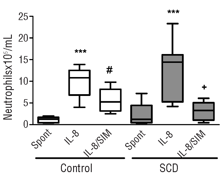
Design and methods: Sickle cell disease patients in steady state and not on hydroxyurea were included in the study. Endothelial cells treated, or not, with tumor necrosis factor-α and simvastatin were used for neutrophil adhesion assays. Neutrophils treated with simvastatin were submitted to interleukin 8-stimulated chemotaxis assays.
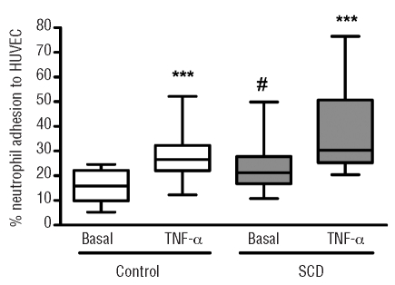
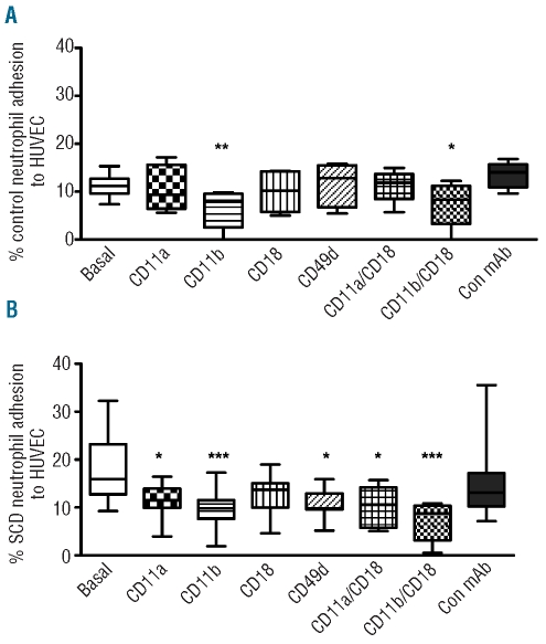
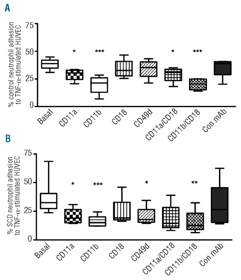
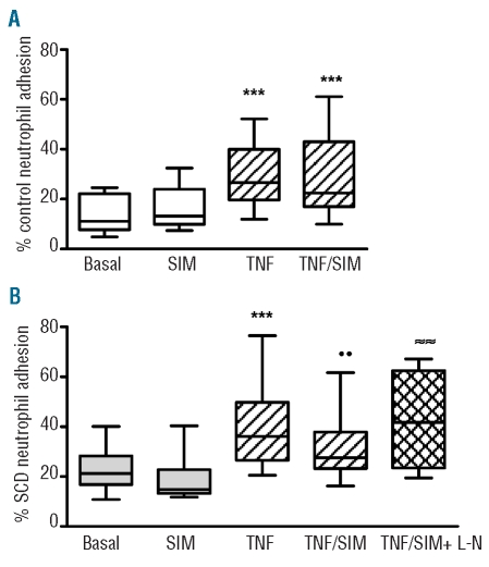
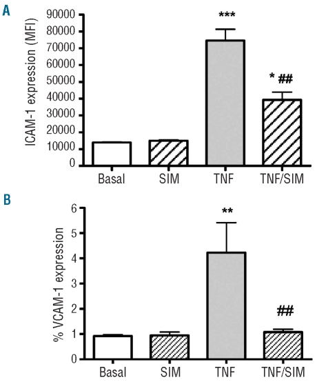
Results: Sickle cell disease neutrophils showed greater adhesion to endothelial cells than control neutrophils. Adhesion of control neutrophils to endothelial cells was mediated by Mac-1 under basal conditions and by the Mac-1 and LFA-1 integrins under inflammatory conditions. In contrast, adhesion of sickle cell disease neutrophils to endothelium, under both basal and tumor necrosis factor-α-stimulated conditions, was mediated by Mac-1 and LFA-1 integrins and also by VLA-4. Under stimulated inflammatory conditions, simvastatin significantly reduced sickle cell disease neutrophil adhesion, and this effect was reversed by inhibition of nitric oxide synthase. Furthermore, intercellular adhesion molecule-1 expression was significantly abrogated on tumor necrosis factor-α-stimulated endothelium incubated with simvastatin, and statin treatment inhibited the interleukin-8-stimulated migration of both control and sickle cell disease neutrophils.
Conclusions: The integrins Mac-1, LFA-1 and, interestingly, VLA-4 mediate the adhesion of sickle cell disease leukocytes to activated endothelial cell layers, in vitro. Our data indicate that simvastatin may be able to reduce endothelial activation and consequent leukocyte adhesion in this in vitro model; future experiments and clinical trials may determine whether simvastatin therapy could be employed in patients with sickle cell disease, with beneficial effects on vaso-occlusion.
Figures
Comment in
-
The rationale for using hydroxycarbamide in the treatment of sickle cell disease.Haematologica. 2011 Apr;96(4):488-91. doi: 10.3324/haematol.2011.041988. Haematologica. 2011. PMID: 21454878 Free PMC article. No abstract available.
References
-
- Okpala I. The intriguing contribution of white blood cells to sickle cell disease - a red cell disorder. Blood Rev. 2004;18(1):65–73. - PubMed
-
- Chiang EY, Frenette PS. Sickle cell vaso-occlusion. Hematol Oncol Clin North Am. 2005;19(5):771–84. - PubMed
-
- Brittain JE, Knoll CM, Ataga KI, Orringer EP, Parise LV. Fibronectin bridges monocytes and reticulocytes via integrin alpha4beta1. Br J Haematol. 2008;141(6):872–81. - PubMed
-
- Fadlon E, Vordermeier S, Pearson TC, Mire-Sluis AR, Dumonde DC, Phillips J, et al. Blood polymorphonuclear leukocytes from the majority of sickle cell patients in the crisis phase of the disease show enhanced adhesion to vascular endothelium and increased expression of CD64. Blood. 1998;91(1):266–74. - PubMed
Publication types
MeSH terms
Substances
LinkOut - more resources
Full Text Sources
Other Literature Sources
Medical
Research Materials
