. 2011 Feb 25;286(8):6733-41.
doi: 10.1074/jbc.M110.175877. Epub 2010 Dec 20.

ATP induces conformational changes in the carboxyl-terminal region of ClC-5

Affiliations

ATP induces conformational changes in the carboxyl-terminal region of ClC-5

Leigh Wellhauser et al. J Biol Chem. .

Abstract

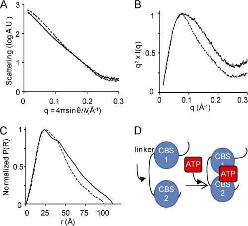
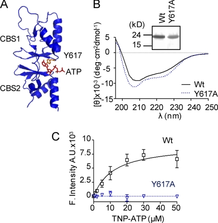
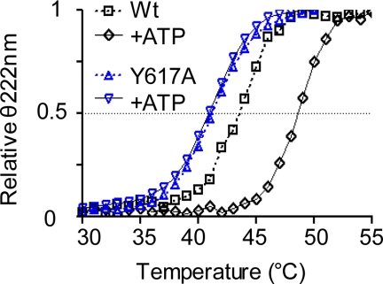
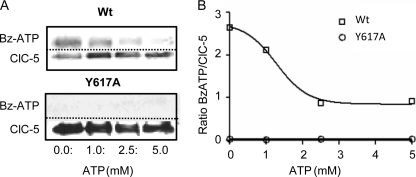
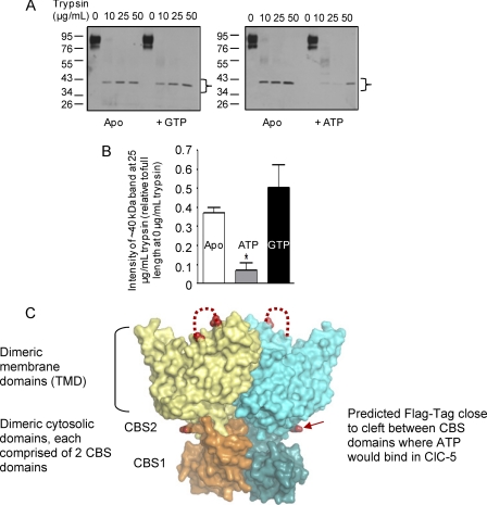
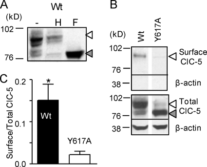
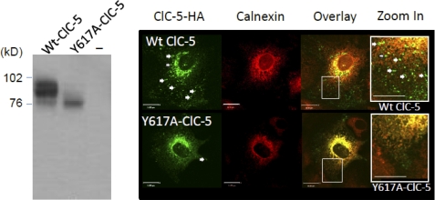
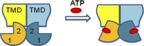
ATP binding enhances the activity of ClC-5, the transporter mutated in Dent disease, a disease affecting the renal proximal tubule. Previously, the ATP binding site was revealed in x-ray crystal structures of the cytoplasmic region of this membrane protein. Disruption of this site by mutagenesis (Y617A-ClC-5) reduced the functional expression and ATP-dependent regulation of the full-length transporter in Xenopus oocytes. However, insight into the conformational changes underlying ATP-dependent regulation is lacking. Here, we show that ATP binding induces a change in protein conformation. Specifically, small angle x-ray scattering experiments indicate that ATP binding promotes a clamp-like closure of the isolated ClC-5 carboxyl-terminal region. Limited proteolysis studies show that ATP binding induces conformational compaction of the carboxyl-terminal region in the intact membrane protein as well. In the context of fibroblasts and proximal tubule epithelial cells, disruption of the ATP binding site in full-length ClC-5 (Y617A-ClC-5) led to a defect in processing and trafficking out of the endoplasmic reticulum. These latter findings account for the decrease in functional expression previously reported for this ATP-binding mutant and prompt future study of a model whereby conformational compaction caused by ATP binding promotes biosynthetic maturation.


Figures

FIGURE 1.
FIGURE 1.
ATP binding induces a conformational change in the ClC-5 Ct region. All SAXS experiments were performed on 400 μm ClC-5 Ct peptide in 100 mm Tris, 500 mm NaCl, 5 mm TCEP, pH 8.0, with and without 5 mm ATP. Three independent trials were preformed on separate protein batches, and a representative experiment is shown in A–D. A, SAXS profiles of apo (solid line) or ATP-bound (dotted line) ClC-5 Ct peptide. B, normalized pairwise distribution functions for apo (solid line) and ATP-bound (dotted line) ClC-5 Ct peptide. C, Kratky plot of apo (solid line) or ATP-bound (dotted line) ClC-5 Ct peptide. D, proposed model of the clamp-like motion of ClC-5 CBS domains around the ATP molecule.
FIGURE 2.
FIGURE 2.
ATP binding to the ClC-5 Ct peptide is impaired by the Y617A mutation. A, interaction with Tyr-617 of ClC-5 Ct peptide (Protein Data Bank ID code 2J9L) with ATP, essential for its binding and co-ordination (10). B, CD spectra of WT and mutant (Y617A) Ct peptides (15 μm each in 5 mm Tris, 500 mm NaCl, pH 8.0). Inset represents silver-stained SDS-PAGE of purified proteins. C, concentration curve of TNP-ATP binding by WT (squares) and Y617A (triangles) Ct peptides (4.3 μm) from 0 to 50 μm TNP-ATP. The Kd for WT Ct peptide is 11.87 ± 5.1 μm TNP-ATP (n = 4 trials each).
FIGURE 3.
FIGURE 3.
Y617A ClC-5 Ct mutant is thermally unstable. Thermal melts of purified WT (11) and Y617A Ct peptides (15 μm) ± 500 μm ATP was monitored by θ222 nm. The Tm values for WT protein (apo = 44.16 ± 0.82 and +ATP = 48.95 ± 0.24 °C, p = 0.001 (11) and Y617A protein (apo = 41.15 ± 0.08 and +ATP 41.37 ± 0.12 °C, p = 0.205) were measured.
FIGURE 4.
FIGURE 4.
Full-length ClC-5 in Sf9 insect cell membranes binds ATP directly. A, photoaffinity labeling of WT or Y617A ClC-5 in Sf9 membrane vesicles (100 μg of total protein) with 50 μm radioactive Bz-ATP without and with MgATP2− (1–5.0 mm). Following labeling ClC-5 was solubilized, and protein was immunoprecipitated (α-HA) and separated on SDS-PAGE and transferred to nitrocellulose. The nitrocellulose was probed using Western blotting (α-HA) to determine the amount of protein present and exposed to autoradiograph film to determine the amount of Bz-ATP labeling. B, quantification of the labeling (autoradiograph) signal versus protein (Western) measured using densitometry (ImageJ). The data obtained for WT ClC-5 (squares) was fitted to a competitive binding algorithm (IC50 = 1.28 ± 0.171 mm ATP, r2 = 0.99). No radioactive labeling was detected for Y617A ClC-5 despite abundant protein present.
FIGURE 5.
FIGURE 5.
ATP binding reduces trypsin susceptibility of Ct region in WT, full-length ClC-5 protein. A, Western blots (α-FLAG) of WT ClC-5 (100 μg of total protein), containing a Ct FLAG tag, incubated without and with 5 mm GTP or ATP for 1 h prior to being subjected to limited trypsin (0–50 μg/ml) proteolysis for 15 min at 4 °C. Representative blots for each condition are shown, and the ∼40 kDa band quantified in B is indicated by a bracket. B, bar graph of trypsin susceptibility of ClC-5 without and with the presence (Apo; open bar) of 5 mm GTP (black filled bar) or ATP (gray filled bar). Specifically, the intensity of the ∼40 kDa band (detected by the α-FLAG antibody) at 25 μg/ml trypsin was quantified for each condition using ImageJ. The abundance of the ∼40 kDa band generated from untreated ClC-5 membranes (0.37 ± 0.03 arbitrary units; n = 4) or membranes treated with GTP (0.50 ± 0.12 arbitrary units; n = 3) was significantly higher than the band intensity generated in the presence of ATP (0.07 ± 0.12, n = 3) at 25 μg/ml trypsin (*, p = 0.001 (versus apo) and p = 0.0164 (versus GTP)). C, size of the proteolytic product of ∼40 kDa in size indicates that trypsin likely cuts ClC-5 in the loop connecting transmembrane α-helices L and M as highlighted (red stippled line) on the structure of C. merolae ClC (Cm-ClC, a eukaryotic ortholog, aligned with ClC-5 by Feng et al. (26), Protein Data Bank ID code 3ORG). Three putative trypsin sites lie in this loop in ClC-5 as predicted by ExPASy. Cm-ClC provides a model for ClC-5. Cm-ClC is a homodimer, with each monomer possessing a TMD and a cytosolic domain with two CBS domains. The extreme carboxyl terminus of Cm-ClC after truncation by seven residues is shown using a red arrow. This terminus lies close to the cleft between the CBS domains, the cleft to which ATP binds in ClC-5 and the membrane domain (12). This image was generated using PyMOL (DeLano Scientific LLC).
FIGURE 6.
FIGURE 6.
Y617A ClC-5 mutant is misprocessed in mammalian cells. A, WT ClC-5 protein expressed in CHO cells was treated with endoglycosidase H (H) or N-glycosidase F (F). Both a complex glycosylated (90–100-kDa proteins, white triangle) and core glycosylated (76–80 kDa, gray triangle) forms are observed. B and C, cell surface biotinylation of WT and Y617A ClC-5 in CHO cells is shown as is quantification of mean pixel intensity of surface versus total ClC-5 expression for WT and Y617A (0.15 ± 0.04, n = 5 and 0.02 ± 0.01, n = 3 surface/total respectively, *, p = 0.005). The fidelity of the biotinylation reaction in labeling cell surface expressed rather than intracellular proteins was confirmed as the lack of actin biotinylation (B, top panel).
FIGURE 7.
FIGURE 7.
ATP-binding mutant (Y617A-ClC-5) exhibits misprocessing and mistrafficking in proximal tubule epithelial cells. Left panel shows immunoblot of lysates from OK cells expressing WT-ClC-5, Y617A-ClC-5 (each bearing an amino-terminal HA tag) or untransfected (−) and subjected to analysis by SDS-PAGE. The upper band indicates complex glycosylated Wt ClC-5 and the lower band, core glycosylated ClC-5 in both Wt-ClC-5 and Y617A-ClC-5. The confocal micrographs show the relative localization of recombinant Wt ClC-5 in the upper panel and Y617A-ClC-5 in the lower panel (labeled green). Cells were also labeled using an anti-calnexin antibody (red images). Zoom-in of the area defined using the white box in the overlay of the two stains revealed a distinct expression pattern for the two different proteins. WT ClC-5 in the biosynthetic compartment co-localizes with calnexin, but there is also Wt ClC-5 protein outside of the ER, expressed in punctate structures. On the other hand, the Y617A-ClC-5 expression pattern overlaps almost completely with that of calnexin. These images are representative of 82 and 66 images for each of the WT and Y617A genotypes, respectively. The scale bars represent 16 μm in the three images on the left and 10 μm for the zoomed in image on the far right.
FIGURE 8.
FIGURE 8.
Model of ATP-dependent conformational change in full-length ClC-5. Enhanced global compactness of the intracellular Ct region of ClC-5 upon ATP binding is shown. The conformational compactness induced by ATP binding to the Ct regions (pie shapes, labeled 1 and 2 to denote CBS1 and CBS2 domains, as shown in our biochemical studies). This compactness of the Ct is also associated with its enhanced interaction with the TMD as suggested in trypsin susceptibility studies. We speculate that this large scale conformational change contributes to the activation of its transporter function as documented previously by Zifarelli and Pusch (6) and its biosynthetic processing as shown in the current paper.

Similar articles

Cited by

References

    1. Lloyd S. E., Pearce S. H., Fisher S. E., Steinmeyer K., Schwappach B., Scheinman S. J., Harding B., Bolino A., Devoto M., Goodyer P., Rigden S. P., Wrong O., Jentsch T. J., Craig I. W., Thakker R. V. (1996) Nature 379, 445–449 - PubMed
    1. Lloyd S. E., Gunther W., Pearce S. H., Thomson A., Bianchi M. L., Bosio M., Craig I. W., Fisher S. E., Scheinman S. J., Wrong O., Jentsch T. J., Thakker R. V. (1997) Hum. Mol. Genet. 6, 1233–1239 - PubMed
    1. Wrong O. M., Norden A. G., Feest T. G. (1994) Q. J. Med. 87, 473–493 - PubMed
    1. Scheel O., Zdebik A. A., Lourdel S., Jentsch T. J. (2005) Nature 436, 424–427 - PubMed
    1. Picollo A., Pusch M. (2005) Nature 436, 420–423 - PubMed

Publication types

MeSH terms

LinkOut - more resources