. 2011 Mar;31(5):1066-75.
doi: 10.1128/MCB.00883-10. Epub 2010 Dec 20.

β-Arrestin 1 inhibits the GTPase-activating protein function of ARHGAP21, promoting activation of RhoA following angiotensin II type 1A receptor stimulation

Affiliations

β-Arrestin 1 inhibits the GTPase-activating protein function of ARHGAP21, promoting activation of RhoA following angiotensin II type 1A receptor stimulation

D F Anthony et al. Mol Cell Biol. 2011 Mar.

Abstract

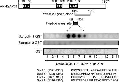
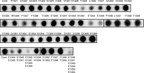
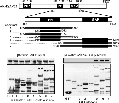
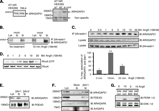
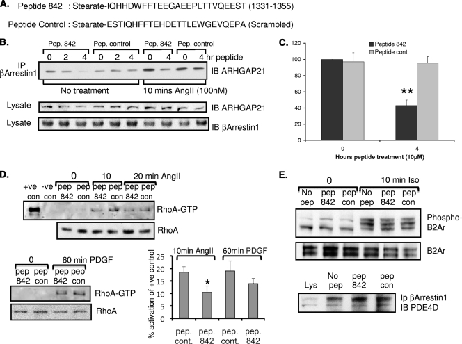
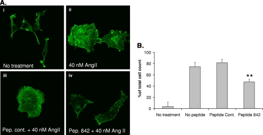
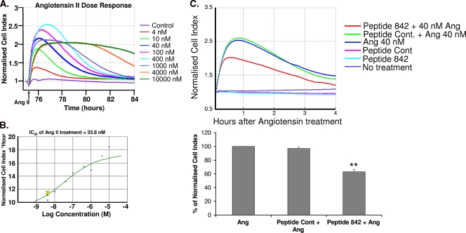
Activation of the small GTPase RhoA following angiotensin II stimulation is known to result in actin reorganization and stress fiber formation. Full activation of RhoA, by angiotensin II, depends on the scaffolding protein β-arrestin 1, although the mechanism behind its involvement remains elusive. Here we uncover a novel partner and function for β-arrestin 1, namely, in binding to ARHGAP21 (also known as ARHGAP10), a known effector of RhoA activity, whose GTPase-activating protein (GAP) function it inhibits. Using yeast two-hybrid screening, a peptide array, in vitro binding studies, truncation analyses, and coimmunoprecipitation techniques, we show that β-arrestin 1 binds directly to ARHGAP21 in a region that transects the RhoA effector GAP domain. Moreover, we show that the level of a complex containing β-arrestin 1 and ARHGAP21 is dynamically increased following angiotensin stimulation and that the kinetics of this interaction modulates the temporal activation of RhoA. Using information gleaned from a peptide array, we developed a cell-permeant peptide that serves to inhibit the interaction of these proteins. Using this peptide, we demonstrate that disruption of the β-arrestin 1/ARHGAP21 complex results in a more active ARHGAP21, leading to less-efficient signaling via the angiotensin II type 1A receptor and, thereby, attenuation of stimulated stress fiber formation.


Figures

FIG. 1.
FIG. 1.
Association of ARHGAP21 and β-arrestin 1 by yeast 2-hybrid and peptide array analyses. (Top) Schematic diagram showing the functionally relevant areas of ARHGAP21. Overlapping sequences within ARHGAP21 that were shown to interact with β-arrestin 1 by yeast 2-hybrid analysis (shaded rectangle) and peptide array (filled rectangle) are indicated. (Bottom) Immobilized peptide spots of overlapping 25-mer peptides, each shifted along by 5 amino acids in the entire sequence of ARHGAP21, probed for interaction with either β-arrestin 1-GST, β-arrestin 2-GST, or GST alone. Positive interactions were detected in amino acid sequences spanning positions 1321 to 1360 of ARHGAP21.
FIG. 2.
FIG. 2.
Alanine scanning arrays of the β-arrestin 1 binding site on ARHGAP21. The alanine substitution array for the P1321-to-P1360 peptide (Cont) (top leftmost spot) from ARHGAP21was probed with β-arrestin 1-GST. A1343 was replaced with an aspartate residue. Multiple substitutions were also undertaken as indicated (bottom).
FIG. 3.
FIG. 3.
β-Arrestin 1 binds to the GAP domain of ARHGAP21. (Top) Purified GST fusions with the indicated regions of the sequence of ARHGAP21 (amino acids 885 to 1346) and GST alone were mixed with purified MBP fusions of β-arrestin 1. (Bottom) Inputs of purified β-arrestin 1-MBP and ARHGAP21-GST constructs (left) and pulled down GST fusion proteins (right) were blotted for the presence of β-arrestin 1-MBP. Asterisks indicate bands that correspond either to an ARHGAP21-GST construct (constructs 1 to 7) or to GST alone (first lane).
FIG. 4.
FIG. 4.
Dynamic interaction between β-arrestin 1 and ARHGAP21 in AT1AR HEK 293 cells. (A) (Left) Cell lysates from AT1AR HEK 293 cells and HeLa cells were blotted for ARHGAP21. (Right) The specificity of the antibody was verified using siRNA silencing of ARHGAP21. cont, control. (B) Immunoprecipitates (IP) of β-arrestin 1 from AT1AR HEK 293 cells were blotted (IB) for ARHGAP21 before or after treatment with angiotensin II (AngII) (100 nM). (C) (Top) Immunoprecipitates of β-arrestin 1 from AT1AR HEK 293 cells were blotted for ARHGAP21 following a time course of treatment with angiotensin II (100 nM). (Center panels) Equal amounts of ARHGAP21 and β-arrestin 1 in inputs. (Bottom) Bar chart showing quantifications of the upper trace. max., maximum; βarr1, β-arrestin 1. (D) RhoA activation in AT1AR HEK 293 cells as measured by a GST-Rhotekin pulldown assay following a time course of angiotensin II treatment. (E) Immunoprecipitates of β-arrestin 1 or β-arrestin 2 from AT1AR HEK 293 cells were blotted for ARHGAP21 and PDE4D. (F) Immunoprecipitates of β-arrestin 1 from AT1AR HEK 293 cells were blotted for ARHGAP21, PDE4D, ARHGAP6, and ArfGAP3. (G) Determination of total and phospho-ERK 1 and 2 in lysates of AT1AR HEK 293 cells following angiotensin II treatment after pretreatment with peptide 842 (pep 842) or a control peptide (pep con). PERK, phospho-ERK.
FIG. 5.
FIG. 5.
Peptide disruption of the β-arrestin 1/ARHGAP21 complex in HEK AT1AR cells. (A) Sequences of the cell-permeant ARHGAP21/β-arrestin 1 disruptor peptide (peptide 842) based on the sequence of ARHGAP21 (amino acids 1331 to 1355) and a scrambled control peptide. (B) (Top) Immunoprecipitates (IP) of β-arrestin 1 from AT1AR HEK 293 cells pretreated with peptide (Pep.) 842 or a peptide control for the indicated times were blotted for ARHGAP21 before or after treatment with angiotensin II (AngII) (100 nM). (Center and bottom) Equal amounts of ARHGAP21 and β-arrestin 1 in inputs. (C) Densitometric analysis of the data shown in the four rightmost lanes of panel B. Results of 3 experiments are shown. **, P < 0.01. (D) (Top) RhoA activation in AT1AR HEK 293 cells as measured by a GST-Rhotekin pulldown assay following a time course of angiotensin II (100 nM) treatment with and without pretreatment (2 h) with peptide 842 or a control (con) peptide. +ve, positive; −ve, negative. (Bottom left) RhoA activation in AT1AR HEK 293 cells as measured by a GST-Rhotekin pulldown assay following PDGF (20 ng/ml) treatment with or without pretreatment (2 h) with peptide 842 or a control peptide. (Bottom right) Quantification by densitometry of the data shown on the left. The results of 3 experiments are shown. *, P < 0.05. (E) (Top and center) Western blot analyses of HEKB2 293 cell lysates following isoprenaline (10 μM) treatment with or without pretreatment (2 h) with peptide 842 or a control peptide. Samples were blotted for the β2-adrenergic receptor (B2Ar) and the phospho-β2-adrenergic receptor (Ser345/346). (Bottom) Immunoprecipitates of β-arrestin 1 from AT1AR HEK 293 cells pretreated with peptide 842 or a peptide control were blotted for PDE4D.
FIG. 6.
FIG. 6.
Disruption of the β-arrestin 1/ARHGAP21 complex affects angiotensin II (AngII)-stimulated stress fiber formation. (A) Visualization of angiotensin II-mediated stress fiber formation in AT1AR HEK 293 cells following pretreatment with peptide (Pep.) 842 or a peptide control (cont.). (B) Analysis of the percentage of total cells containing angiotensin II-mediated stress fibers following pretreatment with peptide 842 or a peptide control. Asterisks indicate a significant difference (P < 0.01) from the peptide control.
FIG. 7.
FIG. 7.
Disruption of the β-arrestin 1/ARHGAP21 complex affects AT1AR signaling. (A) Real-time detection of dose-response curves from AT1AR HEK 293 cells treated with angiotensin II as detected using xCELLigence technology. The arrow indicates the time of addition of angiotensin II (Ang II). (B) Determination of IC50s for the angiotensin II response in AT1AR HEK 293 cells using data from panel A. (C) Evaluation of the effects of pretreatment with peptide 842 or a peptide control (Cont.) on the response to angiotensin II treatment (1 h) in AT1AR HEK 293 cells by using xCELLigence technology. Asterisks indicate a significant difference (P < 0.01) from the value for the peptide control.

References

    1. Altschul, S. F., W. Gish, W. Miller, E. W. Myers, and D. J. Lipman. 1990. Basic local alignment search tool. J. Mol. Biol. 215:403-410. - PubMed
    1. Atienza, J. M., et al. 2006. Dynamic and label-free cell-based assays using the real-time cell electronic sensing system. Assay Drug Dev. Technol. 4:597-607. - PubMed
    1. Baillie, G. S., et al. 2007. Mapping binding sites for the PDE4D5 cAMP-specific phosphodiesterase to the N- and C-domains of β-arrestin using spot-immobilized peptide arrays. Biochem. J. 404:71-80. - PMC - PubMed
    1. Barnes, W. G., et al. 2005. β-Arrestin 1 and Gαq/11 coordinately activate RhoA and stress fiber formation following receptor stimulation. J. Biol. Chem. 280:8041-8050. - PubMed
    1. Bassères, D. S., E. V. Tizzei, A. A. Duarte, F. F. Costa, and S. T. Saad. 2002. ARHGAP10, a novel human gene coding for a potentially cytoskeletal Rho-GTPase activating protein. Biochem. Biophys. Res. Commun. 294:579-585. - PubMed

Publication types

MeSH terms

LinkOut - more resources