. 2011 Jan 4;108(1):137-42.
doi: 10.1073/pnas.1013275108. Epub 2010 Dec 20.

Intracellular organelles mediate cytoplasmic pulling force for centrosome centration in the Caenorhabditis elegans early embryo

Affiliations

Intracellular organelles mediate cytoplasmic pulling force for centrosome centration in the Caenorhabditis elegans early embryo

Kenji Kimura et al. Proc Natl Acad Sci U S A. .

Abstract

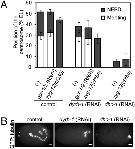
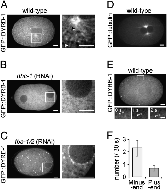
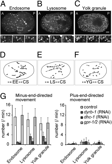
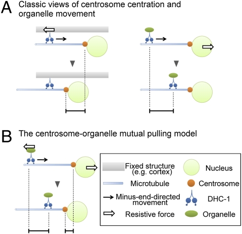
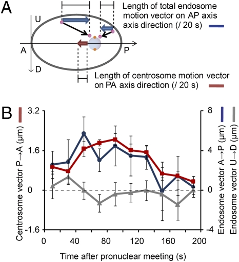
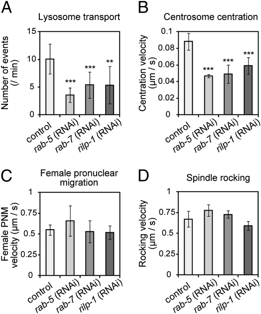
The centrosome is generally maintained at the center of the cell. In animal cells, centrosome centration is powered by the pulling force of microtubules, which is dependent on cytoplasmic dynein. However, it is unclear how dynein brings the centrosome to the cell center, i.e., which structure inside the cell functions as a substrate to anchor dynein. Here, we provide evidence that a population of dynein, which is located on intracellular organelles and is responsible for organelle transport toward the centrosome, generates the force required for centrosome centration in Caenorhabditis elegans embryos. By using the database of full-genome RNAi in C. elegans, we identified dyrb-1, a dynein light chain subunit, as a potential subunit involved in dynein anchoring for centrosome centration. DYRB-1 is required for organelle movement toward the minus end of the microtubules. The temporal correlation between centrosome centration and the net movement of organelle transport was found to be significant. Centrosome centration was impaired when Rab7 and RILP, which mediate the association between organelles and dynein in mammalian cells, were knocked down. These results indicate that minus end-directed transport of intracellular organelles along the microtubules is required for centrosome centration in C. elegans embryos. On the basis of this finding, we propose a model in which the reaction forces of organelle transport generated along microtubules act as a driving force that pulls the centrosomes toward the cell center. This is the first model, to our knowledge, providing a mechanical basis for cytoplasmic pulling force for centrosome centration.


Conflict of interest statement

The authors declare no conflict of interest.

Figures

Fig. 1.
Fig. 1.
DYRB-1 is required for centrosome centration. (A) Mean position of the centrosomes at pronuclear meeting (white bars) and nuclear envelope breakdown (NEBD, gray bars) under the indicated conditions are shown. In dhc-1 (RNAi) embryos or the zyg-12(ct350) background at restrictive temperature, pronuclear meeting did not occur. Position is expressed as percentage of EL (posterior-most side of the egg, 0%). The average value ± SD is shown (n ≥ 5). (B) Time-lapse images of control (untreated), dyrb-1 (RNAi), and dhc-1 (RNAi) embryos expressing GFP-tubulin under the zyg-12(ct350) background at restrictive temperature were projected onto single images to visualize the trajectory of the centrosomes movement until NEBD. (Scale bars, 5 μm.)
Fig. 2.
Fig. 2.
Cytoplasmic localization of GFP::DYRB-1 during centrosome centration. Confocal fluorescence images of C. elegans embryos expressing GFP::DYRB-1 are shown (AC). The boxed region in each panel is also shown magnified (Right). The arrowheads (black and white) in A indicate both ends of a filamentous signal of GFP::DYRB-1: (B) dhc-1 (RNAi) and (C) tba-1/2 (RNAi). (D) The pattern of astral microtubules revealed using a GFP::tubulin strain. (E) Tracking of DYRB-1 puncta. A fraction of DYRB-1 shows a punctate signal, a portion of which moves directionally as shown in the lower magnified images (bar, 2 μm). (F) The direction and frequency of fast (≥0.5 μm/s) and continuous (≥2 s) movements of DYRB-1 puncta within 30 s after pronuclear meeting (n = 10). The average number ± SEM is shown. (Scale bars, 5 μm except in E, Lower.)
Fig. 3.
Fig. 3.
Movements of intracellular organelles during centrosome centration. Confocal fluorescence images of (A) an embryo expressing both GFP::TBG-1 (arrow) and GFP::EEA-1(FYVE*2), (B) an embryo in which the lysosomes were stained with LysoTracker, and (C) an embryo expressing the yolk granule marker GFP::VIT-2. A, anterior; P, posterior. (Scale bar, 5 μm.) Three sequential fluorescence images of the boxed regions in AC are magnified (bar, 2 μm) below each panel. The arrowhead marks a moving organelle. (DF) A typical example of the movements of early endosomes (EE), lysosomes (LS), or yolk granules (YG; ≥0.5 μm/s, black arrows) for 2 min after pronuclear meeting is illustrated. Black ellipse represents the outline of the egg; gray arrows mark the movement of centrosomes (CS). (G) The number of each organelle that moved continuously for at least 2 s at a rate of at least 0.5 μm/s toward the minus end or plus end of the microtubules was counted for 2 min after pronuclear meeting in control, dyrb-1 (RNAi), dhc-1 (RNAi), and gpr-1/2 (RNAi) embryos (n ≥ 5). The average number ± SD of each organelle per 1 min is shown.
Fig. 4.
Fig. 4.
The centrosome–organelle mutual pulling model for centrosome centration. (A) Classic views of centrosome centration and organelle movement in which either dynein (Left) or the centrosomes (Right) are fixed and the other side (centrosomes or organelles, respectively) moves. (B) The centrosome–organelle mutual pulling model. When dyneins slide along the microtubule, both dynein–organelle complexes and the centrosomes move toward each other. This mutual movement positions the centrosomes at the cell center.
Fig. 5.
Fig. 5.
The correlation between early endosome movement and centrosome centration. (A) Schematic of the measurement of the movement of early endosomes [GFP::EEA-1(FYVE*2), blue arrows] and centrosomes (GFP::TBG-1, red arrow) along the AP axis. The net movements of early endosomes in the anterior (A)-to-posterior (P) direction were calculated by subtracting the sum of P-to-A movements from the sum of A-to-P movements. Likewise, the net movements of early endosomes in the perpendicular direction to the AP axis [upside (U) – downside (D)] were calculated. (Gray ellipse, embryo; light blue circle, pronucleus; pink circle, early endosome; black arrow, movement of early endosome.) The length of movement of total early endosomes and the centrosomes was measured during a 200-s period just after pronuclear meeting. (B) The average length of movement of total early endosomes (blue line, AP axis component; gray line, perpendicular component) and centrosomes (red line, AP axis component) during each 20-s period on the equatorial plane is plotted (n = 9). Left y axis, the length of the centrosome movements (μm); right y axis, the length of total early endosome movements (μm); x axis, time of the measurement from after pronuclear meeting. Error bars represent SEM.
Fig. 6.
Fig. 6.
Centrosome centration was delayed when organelle movement was impaired. (A) The average number ± SD of lysosomes that moved continuously for at least 2 s at a rate of at least 0.5 μm/s during the 60 s after pronuclear meeting in WT, rab-5 (RNAi), rab-7 (RNAi), and rilp-1 (RNAi) embryos (n ≥ 7). (B) Average velocity ± SD (μm/s) of centrosome centration (n ≥ 7). (C) Average velocity ± SD (μm/s) of female pronuclear migration during the fast phase (n ≥ 8). (D) Average velocity ± SD (μm/s) of the maximum swing of the posterior spindle pole at metaphase (n ≥ 4). A statistically significant difference from control is indicated by asterisks (***P < 0.001, **P < 0.01).

References

    1. Howard J, Hyman AA. Dynamics and mechanics of the microtubule plus end. Nature. 2003;422:753–758. - PubMed
    1. Rios RM, Bornens M. The Golgi apparatus at the cell centre. Curr Opin Cell Biol. 2003;15:60–66. - PubMed
    1. Doxsey S, McCollum D, Theurkauf W. Centrosomes in cellular regulation. Annu Rev Cell Dev Biol. 2005;21:411–434. - PubMed
    1. Nigon V, Guerrier P, Monin H. L'architecture polaire de l'oeuf et les mouvements des constituants cellulaires au cours des premieres etapes du developpement chez quelques nematodes. Bull Biol Fr Belg. 1960;93:131–202.
    1. Albertson DG. Formation of the first cleavage spindle in nematode embryos. Dev Biol. 1984;101:61–72. - PubMed

Publication types

MeSH terms

LinkOut - more resources