. 2011 Jan 4;108(1):266-71.
doi: 10.1073/pnas.1016791108. Epub 2010 Dec 20.

Single dose of anti-CTLA-4 enhances CD8+ T-cell memory formation, function, and maintenance

Affiliations

Single dose of anti-CTLA-4 enhances CD8+ T-cell memory formation, function, and maintenance

Virginia A Pedicord et al. Proc Natl Acad Sci U S A. .

Abstract

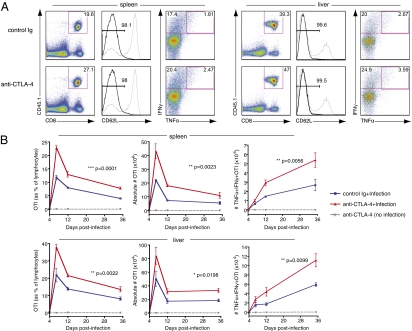
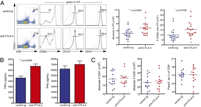
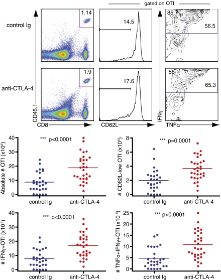
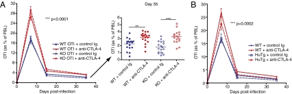
CTLA-4, an Ig superfamily molecule with homology to CD28, is one of the most potent negative regulators of T-cell responses. In vivo blockade of CTLA-4 exacerbates autoimmunity, enhances tumor-specific T-cell responses, and may inhibit the induction of T-cell anergy. Clinical trials of CTLA-4-blocking antibodies to augment T-cell responses to malignant melanoma are at an advanced stage; however, little is known about the effects of CTLA-4 blockade on memory CD8(+) T-cell responses and the formation and maintenance of long-term CD8(+) T-cell memory. In our studies, we show that during in vivo memory CD8(+) T-cell responses to Listeria monocytogenes infection, CTLA-4 blockade enhances bacterial clearance and increases memory CD8(+) T-cell expansion. This is followed by an accumulation of memory cells that are capable of producing the effector cytokines IFN-γ and TNF-α. We also demonstrate that in a vaccination setting, blocking CTLA-4 during CD8(+) T-cell priming leads to increased expansion and maintenance of antigen-specific memory CD8(+) T cells without adversely affecting the overall T-cell repertoire. This leads to an increase in memory cell effector function and improved protective immunity against further bacterial challenges. These results indicate that transient blockade of CTLA-4 enhances memory CD8(+) T-cell responses and support the possible use of CTLA-4-blocking antibodies during vaccination to augment memory formation and maintenance.


Conflict of interest statement

Conflict of interest statement: J.P.A. is a paid consultant for Bristol-Myers Squibb and is the primary inventor on the patent “Blockade of T lymphocyte down-regulation associated with CTLA-4 signaling.”

Figures

Fig. 1.
Fig. 1.
Blockade of CTLA-4 during memory responses increases memory CD8+ T-cell expansion and effector function. Memory OTI cells generated in vivo were sorted and adoptively transferred into new naïve recipients. Recipient mice then received either control Ig or anti–CTLA-4 and an i.v. infection with LM-OVA. (A) Flow cytometry of lymphocytes (left column) and gated CD8+CD45.1+ memory OTI cells (right two columns) in spleen and liver at 7 d after infection. Numbers in gates indicate frequency of OTI as a percentage of lymphocytes (left column) and frequency of CD62L-low and IFNγ+ and double-positive, TNFα+ IFNγ+, as a percentage of gated OTI (middle and right columns, respectively). Gray histograms represent expression levels in total, unfractionated lymphocytes for reference. Each plot is from one mouse (median for each group) of three. (B) Frequency and absolute number of OTI memory cells and effector cytokine-producing OTI memory cells at days 4, 7, 12, and 35 after infection. Data shown are of three mice per group for each time point, and error bars indicate means ± SEM. Two-way ANOVA was used to evaluate statistical significance. Data shown are representative of three independent experiments.
Fig. 2.
Fig. 2.
Presence of anti–CTLA-4 during priming increases memory cells and their effector function without adversely affecting overall T-cell repertoire. Naïve OTI cells were adoptively transferred into B6 recipients that were then injected with either control Ig or anti–CTLA-4 just before infection with LM-OVA. Spleens were analyzed at least 45 d after infection, with no further treatment or antigen exposure. (A) Left: Flow cytometry of lymphocytes (left column) and gated CD8+CD45.1+ memory OTI cells (right three columns) from spleens of mice that received either control Ig or anti–CTLA-4 during priming. Numbers in gates indicate frequency of OTI as a percentage of lymphocytes (left column) and frequency of CD62L-low, CD127+, and CD44-high as percentage of gated OTI (right three columns). Gray histograms represent expression levels in total, unfractionated lymphocytes for reference. Each plot is from one mouse (median for each group) of seven. Right: Graphs of quantified data pooled from three independent experiments with six to seven mice per group. (B) Cells were restimulated with SIINFEKL for 20 h, and supernatants were analyzed for effector cytokine secretion using cytometric bead arrays. Pooled data from three independent experiments are shown, and error bars indicate means ± SEM. (C) Absolute numbers of CD4+ and CD8+ T cells and frequencies of Tregs from spleens of treated mice were quantified from flow cytometry. Data shown are pooled from two independent experiments with six to seven mice per group. Statistical significance was determined with t tests.
Fig. 3.
Fig. 3.
Blockade of CTLA-4 during priming enhances memory CD8+ T-cell effector function. Naïve OTI cells were adoptively transferred into B6 recipients and injected with either control Ig or anti–CTLA-4 just before infection with LM-OVA. After at least 60 d, mice received a rechallenge infection with LM-OVA. Upper: Flow cytometry of lymphocytes (left column) and gated CD8+CD45.1+ memory OTI cells (right two columns) in spleens at 24 h after infection. Numbers in gates indicate frequency of OTI as a percentage of lymphocytes (left column) and frequency of CD62L-low and IFNγ+ and double-positive, TNFα+ IFNγ+ as a percentage of gated OTI (middle and right columns, respectively). Each plot is from 1 mouse (median) of 10. Lower: Graphs of quantified data pooled from three independent experiments with 9 to 10 mice per group. Statistical significance was determined with t tests.
Fig. 4.
Fig. 4.
Anti–CTLA-4 enhancement of memory CD8+ T cells requires cell-extrinsic blockade of CTLA-4. (A) Naïve OTI cells from either wild-type (WT) or CTLA-4–deficient (KO) mice were adoptively transferred into new naïve wild-type recipients. Recipient mice then received either control Ig or anti–CTLA-4 and an i.v. infection with LM-OVA. Left: Frequency of antigen-specific OTI cells collected from peripheral blood at days 7, 15, and 35 after infection. Right: Dot plots of OTI frequency in individual mice at day 35. Data shown are pooled from three independent experiments with five to six mice per group. Two-way ANOVA was used to evaluate statistical significance for all time points, and error bars indicate means ± SEM. One-way ANOVA was used for day-35 comparisons. **P < 0.01; ***P < 0.001. (B) Naïve OTI cells were adoptively transferred into naïve recipients, either wild-type or transgenic mice engineered to express only human CTLA-4 (HuTg). Recipient mice then received either control Ig or anti–CTLA-4 and an i.v. infection with LM-OVA, allowing for blockade of CTLA-4 only on cells expressing mouse CTLA-4. Plot shows frequency of OTI cells collected from peripheral blood at days 7, 15, and 35 after infection. Data shown are pooled from two independent experiments with five mice per group and analyzed by two-way ANOVA.

References

    1. Krummel MF, Allison JP. CTLA-4 engagement inhibits IL-2 accumulation and cell cycle progression upon activation of resting T cells. J Exp Med. 1996;183:2533–2540. - PMC - PubMed
    1. Walunas TL, et al. CTLA-4 can function as a negative regulator of T cell activation. Immunity. 1994;1:405–413. - PubMed
    1. Tivol EA, et al. Loss of CTLA-4 leads to massive lymphoproliferation and fatal multiorgan tissue destruction, revealing a critical negative regulatory role of CTLA-4. Immunity. 1995;3:541–547. - PubMed
    1. Waterhouse P, et al. Lymphoproliferative disorders with early lethality in mice deficient in Ctla-4. Science. 1995;270:985–988. - PubMed
    1. Alegre ML, Frauwirth KA, Thompson CB. T-cell regulation by CD28 and CTLA-4. Nat Rev Immunol. 2001;1:220–228. - PubMed

Publication types