

Essential role for mast cell tryptase in acute experimental colitis
- PMID: 21173247
- PMCID: PMC3017166
- DOI: 10.1073/pnas.1005758108
Essential role for mast cell tryptase in acute experimental colitis
Abstract
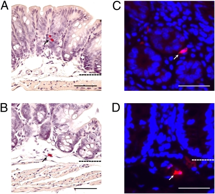
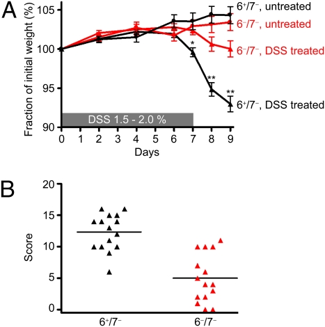
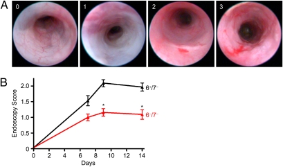
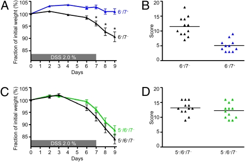
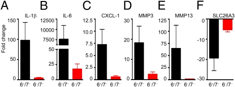
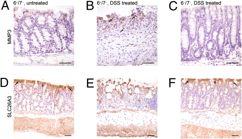
Patients with inflammatory bowel disease (IBD) have increased numbers of human tryptase-β (hTryptase-β)-positive mast cells (MCs) in the gastrointestinal tract. The amino acid sequence of mouse mast cell protease (mMCP)-6 is most similar to that of hTryptase-β. We therefore hypothesized that this mMCP, or the related tryptase mMCP-7, might have a prominent proinflammatory role in experimental colitis. The dextran sodium sulfate (DSS) and trinitrobenzene sulfonic acid (TNBS) colitis models were used to evaluate the differences between C57BL/6 (B6) mouse lines that differ in their expression of mMCP-6 and mMCP-7 with regard to weight loss, colon histopathology, and endoscopy scores. Microarray analyses were performed, and confirmatory real-time PCR, ELISA, and/or immunohistochemical analyses were carried out on a number of differentially expressed cytokines, chemokines, and matrix metalloproteinases (MMPs). The mMCP-6-null mice that had been exposed to DSS had significantly less weight loss as well as significantly lower pathology and endoscopy scores than similarly treated mMCP-6-expressing mice. This difference in colitis severity was confirmed endoscopically in the TNBS-treated mice. Evaluation of the distal colon segments revealed that numerous proinflammatory cytokines, chemokines that preferentially attract neutrophils, and MMPs that participate in the remodeling of the ECM were all markedly increased in the colons of DSS-treated WT mice relative to untreated WT mice and DSS-treated mMCP-6-null mice. Collectively, our data show that mMCP-6 (but not mMCP-7) is an essential MC-restricted mediator in chemically induced colitis and that this tryptase acts upstream of many of the factors implicated in IBD.
Conflict of interest statement
Conflict of interest statement: G.D.L. is an employee of Biomodels, LLC. This company did not fund the study, and the other authors have no affiliation with the company.
Figures

Similar articles
-
Importance of mast cell Prss31/transmembrane tryptase/tryptase-γ in lung function and experimental chronic obstructive pulmonary disease and colitis.J Biol Chem. 2014 Jun 27;289(26):18214-27. doi: 10.1074/jbc.M114.548594. Epub 2014 May 12. J Biol Chem. 2014. PMID: 24821729 Free PMC article.
-
Mast cell-restricted, tetramer-forming tryptases induce aggrecanolysis in articular cartilage by activating matrix metalloproteinase-3 and -13 zymogens.J Immunol. 2013 Aug 1;191(3):1404-12. doi: 10.4049/jimmunol.1300856. Epub 2013 Jun 24. J Immunol. 2013. PMID: 23797671 Free PMC article.
-
The mouse mast cell-restricted tetramer-forming tryptases mouse mast cell protease 6 and mouse mast cell protease 7 are critical mediators in inflammatory arthritis.Arthritis Rheum. 2008 Aug;58(8):2338-46. doi: 10.1002/art.23639. Arthritis Rheum. 2008. PMID: 18668540
-
Protease-proteoglycan complexes of mouse and human mast cells and importance of their beta-tryptase-heparin complexes in inflammation and innate immunity.Immunol Rev. 2007 Jun;217:155-67. doi: 10.1111/j.1600-065X.2007.00525.x. Immunol Rev. 2007. PMID: 17498058 Review.
-
Dextran sodium sulfate colitis murine model: An indispensable tool for advancing our understanding of inflammatory bowel diseases pathogenesis.World J Gastroenterol. 2017 Sep 7;23(33):6016-6029. doi: 10.3748/wjg.v23.i33.6016. World J Gastroenterol. 2017. PMID: 28970718 Free PMC article. Review.
Cited by
-
Mast cell proteases as pharmacological targets.Eur J Pharmacol. 2016 May 5;778:44-55. doi: 10.1016/j.ejphar.2015.04.045. Epub 2015 May 7. Eur J Pharmacol. 2016. PMID: 25958181 Free PMC article. Review.
-
Role of perivascular nerve and sensory neurotransmitter dysfunction in inflammatory bowel disease.Am J Physiol Heart Circ Physiol. 2021 May 1;320(5):H1887-H1902. doi: 10.1152/ajpheart.00037.2021. Epub 2021 Mar 12. Am J Physiol Heart Circ Physiol. 2021. PMID: 33710922 Free PMC article.
-
The Effect of Serine Protease Inhibitors on Visceral Pain in Different Rodent Models With an Intestinal Insult.Front Pharmacol. 2022 Jun 2;13:765744. doi: 10.3389/fphar.2022.765744. eCollection 2022. Front Pharmacol. 2022. PMID: 35721192 Free PMC article.
-
Nuclear receptor 4a3 (nr4a3) regulates murine mast cell responses and granule content.PLoS One. 2014 Feb 20;9(2):e89311. doi: 10.1371/journal.pone.0089311. eCollection 2014. PLoS One. 2014. PMID: 24586680 Free PMC article.
-
Cinnamaldehyde is the main mediator of cinnamon extract in mast cell inhibition.Eur J Nutr. 2015 Dec;54(8):1297-309. doi: 10.1007/s00394-014-0810-0. Epub 2014 Dec 11. Eur J Nutr. 2015. PMID: 25504111
References
-
- Reynolds DS, Gurley DS, Austen KF, Serafin WE. Cloning of the cDNA and gene of mouse mast cell protease-6. Transcription by progenitor mast cells and mast cells of the connective tissue subclass. J Biol Chem. 1991;266:3847–3853. - PubMed
-
- Hunt JE, et al. Natural disruption of the mouse mast cell protease 7 gene in the C57BL/6 mouse. J Biol Chem. 1996;271:2851–2855. - PubMed
-
- Thakurdas SM, et al. The mast cell-restricted tryptase mMCP-6 has a critical immunoprotective role in bacterial infections. J Biol Chem. 2007;282:20809–20815. - PubMed
Publication types
MeSH terms
Substances
Grants and funding
LinkOut - more resources
Full Text Sources
Molecular Biology Databases
