. 2011 Jan 4;108(1):415-20.
doi: 10.1073/pnas.1012076108. Epub 2010 Dec 20.

Mechanism of RNA stabilization and translational activation by a pentatricopeptide repeat protein

Affiliations

Mechanism of RNA stabilization and translational activation by a pentatricopeptide repeat protein

Jana Prikryl et al. Proc Natl Acad Sci U S A. .

Abstract

Pentatricopeptide repeat (PPR) proteins comprise a large family of helical repeat proteins that bind RNA and modulate organellar RNA metabolism. The mechanisms underlying the functions attributed to PPR proteins are unknown. We describe in vitro studies of the maize protein PPR10 that clarify how PPR10 modulates the stability and translation of specific chloroplast mRNAs. We show that recombinant PPR10 bound to its native binding site in the chloroplast atpI-atpH intergenic region (i) blocks both 5'→3' and 3'→ 5 exoribonucleases in vitro; (ii) is sufficient to define the native processed atpH mRNA 5'-terminus in conjunction with a generic 5'→3' exoribonuclease; and (iii) remodels the structure of the atpH ribosome-binding site in a manner that can account for PPR10's ability to enhance atpH translation. In addition, we show that the minimal PPR10-binding site spans 17 nt. We propose that the site-specific barrier and RNA remodeling activities of PPR10 are a consequence of its unusually long, high-affinity interface with single-stranded RNA, that this interface provides a functional mimic to bacterial small RNAs, and that analogous activities underlie many of the biological functions that have been attributed to PPR proteins.


Conflict of interest statement

The authors declare no conflict of interest.

Figures

Fig. 1.
Fig. 1.
Fine-mapping PPR10’s atpH-binding site. (A) The atpI–atpH intergenic region. The sequence shown previously to harbor the PPR10-binding site (5) is in bold, and the minimal binding site defined here is shaded in gray. The RNA termini that are stabilized by PPR10 in vivo are shown below. (B) Gel mobility shift assays were performed with the synthetic RNAs diagrammed below. RNA 4, highlighted in bold, has the minimal sequence that binds with high affinity to PPR10. Binding reactions contained RNAs at 40 pM and rPPR10 at the following concentrations: first and fourth panels: 0, 50, 100, and 200 nM; second panel: 0, 12.5, 25, and 50 nM; third panel: 0, 5, 10, and 20 nM. B, bound; U, unbound. (C) Equilibrium Kd of PPR10 for the minimal atpH-binding site. Binding reactions contained RNA 4 (panel B). The PPR10 concentrations and fraction of RNA bound in each lane are plotted below. The Kd calculation assumed a 1:1 interaction between the RNA and protein. (D) Alignment of the atpH 5′ UTR from Nicotiana tabacum (N. tab), Arabidopsis thaliana (A. tha), and Zea mays (Z. mays). The PPR10-dependent 5′ and 3′ RNA termini and the putative Shine–Dalgarno (SD) element are indicated.
Fig. 2.
Fig. 2.
Both sequence and spacing contribute to the differing affinity of PPR10 for the atpH- and psaJ-binding sites. The wild-type atpH site was assayed in the context of a 29-mer, 19-mer, and 17-mer, as indicated. The minimal PPR10-binding site is shaded. The psaJ oligomer is the same oligomer shown previously to bind PPR10 in vitro (5). Residues that differ from the corresponding positions in the minimal atpH site are underlined. rPPR10 was present at 0, 50, 100, and 200 nM (Left) or 0, 85, and 175 nM (Right).
Fig. 3.
Fig. 3.
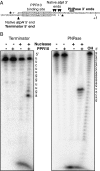
PPR10 blocks 5′→3′ and 3′→5′ exoribonucleases in vitro. (A) RNAs used for the exonuclease protection assays. The RNA at top was labeled at its 5′ end (*) and was used for the PNPase assay. The RNA at bottom was labeled at its 3′ (*) end and was used for the Terminator assay. The PPR10-dependent RNA termini detected in vivo and in vitro are marked. The nucleotide marked “+1” is the first nucleotide of the atpH ORF. (B) PPR10-mediated protection of RNA from Terminator 5′→3′ exonuclease (Left) and PNPase 3′→5′ exonuclease (Right). The Terminator and PNPase reactions used the 3′-end–labeled and 5′-end–labeled RNAs shown in A, respectively. Details of the assay conditions are provided in SI Materials and Methods. The position of each protected terminus was determined from the size of the corresponding end-labeled RNA, which could be determined to single-nucleotide resolution as follows. Terminator generated a ladder of labeled RNAs that share the same 3′ end but whose 5′ ends differ by 1 nt (last lane). The 5′ nucleotide of the RNA in each band is shown in the sequence to the right. For the PNPase experiment, a partial alkaline hydrolysis ladder (OH) of the 5′-end–labeled RNA served the analogous purpose: the 3′ nucleotide of the labeled RNA at each position was determined by its difference in size from the full-length RNA and is shown to the right.
Fig. 4.
Fig. 4.
PPR10 releases the atpH ribosome-binding region from an RNA duplex. (A) RNA (shown in B) was radiolabeled at its 5′ end and incubated with RNase T1 or V1 in the presence or absence of rPPR10. The ribonucleases were used at two different concentrations, as indicated. An alkali hydrolysis ladder (OH) and the RNase T1 cleavage pattern of the denatured RNA (T1) provide size markers. Details of the assay conditions are provided in SI Materials and Methods. (B) Secondary structure of the atpH 5′ UTR and summary of the structure probing data. The RNA structure was predicted by M-fold and is supported by the data in A. The minimal PPR10-binding site is shaded. The atpH start codon and putative Shine–Dalgarno (SD) element are marked. (+) and (−) indicate increased or decreased cleavage, respectively, in the presence of rPPR10. (C) Model for translational activation by PPR10. PPR10 captures its binding site in single-stranded form, releasing the atpH ribosome-binding region from a sequestering RNA structure.

References

    1. Schmitz-Linneweber C, Small I. Pentatricopeptide repeat proteins: A socket set for organelle gene expression. Trends Plant Sci. 2008;13:663–670. - PubMed
    1. Small ID, Peeters N. The PPR motif—a TPR-related motif prevalent in plant organellar proteins. Trends Biochem Sci. 2000;25:46–47. - PubMed
    1. Schmitz-Linneweber C, Williams-Carrier R, Barkan A. RNA immunoprecipitation and microarray analysis show a chloroplast pentatricopeptide repeat protein to be associated with the 5′ region of mRNAs whose translation it activates. Plant Cell. 2005;17:2791–2804. - PMC - PubMed
    1. Schmitz-Linneweber C, et al. A pentatricopeptide repeat protein facilitates the trans-splicing of the maize chloroplast rps12 pre-mRNA. Plant Cell. 2006;18:2650–2663. - PMC - PubMed
    1. Pfalz J, Bayraktar OA, Prikryl J, Barkan A. Site-specific binding of a PPR protein defines and stabilizes 5′ and 3′ mRNA termini in chloroplasts. EMBO J. 2009;28:2042–2052. - PMC - PubMed

Publication types

MeSH terms

Substances