. 2011 Jan 4;108(1):126-30.
doi: 10.1073/pnas.1015032108. Epub 2010 Dec 20.

Experimental support for the evolution of symmetric protein architecture from a simple peptide motif

Affiliations

Experimental support for the evolution of symmetric protein architecture from a simple peptide motif

Jihun Lee et al. Proc Natl Acad Sci U S A. .

Abstract

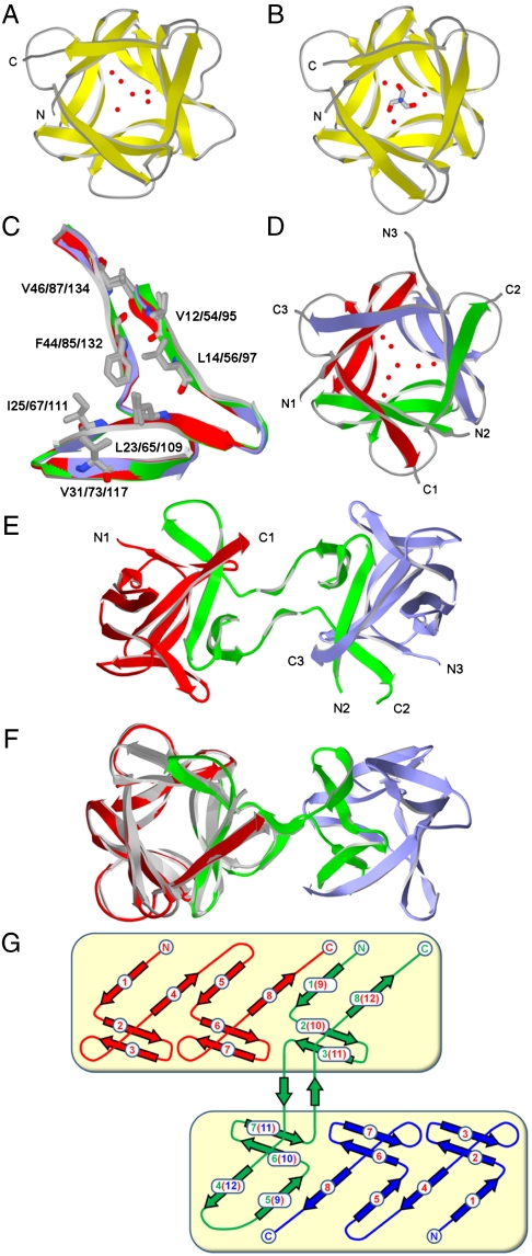
The majority of protein architectures exhibit elements of structural symmetry, and "gene duplication and fusion" is the evolutionary mechanism generally hypothesized to be responsible for their emergence from simple peptide motifs. Despite the central importance of the gene duplication and fusion hypothesis, experimental support for a plausible evolutionary pathway for a specific protein architecture has yet to be effectively demonstrated. To address this question, a unique "top-down symmetric deconstruction" strategy was utilized to successfully identify a simple peptide motif capable of recapitulating, via gene duplication and fusion processes, a symmetric protein architecture (the threefold symmetric β-trefoil fold). The folding properties of intermediary forms in this deconstruction agree precisely with a previously proposed "conserved architecture" model for symmetric protein evolution. Furthermore, a route through foldable sequence-space between the simple peptide motif and extant protein fold is demonstrated. These results provide compelling experimental support for a plausible evolutionary pathway of symmetric protein architecture via gene duplication and fusion processes.


Conflict of interest statement

The authors declare no conflict of interest.

Figures

Fig. 1.
Fig. 1.
Evolutionary models of symmetry protein architecture (e.g., the β-trefoil fold). (A) The emergent architecture model (8). The archaic peptide motif is autonomously folding, yielding simple single-domain architecture. Intermediate forms produced by gene duplication and fusion events have unique folds with increasing structural symmetry and complexity. The symmetric target architecture emerges upon the final gene duplication and fusion event. (B) The conserved architecture model (9). The archaic peptide is not an autonomously folding single-domain architecture; instead, it oligomerizes to yield the symmetric target architecture (e.g., β-trefoil fold). Intermediate forms produced by gene duplication and fusion events similarly oligomerize and reconstitute integral units of the symmetric target architecture. A final duplication and fusion event encodes the symmetric target architecture within a single polypeptide chain.
Fig. 2.
Fig. 2.
Summary of the top-down symmetric deconstruction strategy and the result of its application to FGF-1. (A) The cumulative symmetric transforms applied to achieve a symmetric deconstruction of the β-trefoil architecture (beginning with FGF-1 and ending in the Monofoil and Difoil polypeptides). Details for the set of intermediary mutants comprising the deconstruction are provided in Table S1. (B) Primary structures of the FGF-1 starting protein and the resulting Symfoil-4P symmetric deconstruction. Positions of exact threefold primary structure symmetry are indicated in yellow. The Monofoil-4P and Difoil-4P polypeptides were generated by the introduction of stop codons at positions 53 and 94 of the Symfoil-4P protein, respectively.
Fig. 3.
Fig. 3.
X-ray structures of FGF-1, Symfoil-1, Monofoil-4P, and Difoil-4P proteins. (A) Ribbon representation of FGF-1 oriented down the threefold axis of symmetry and including select solvent structure. (B) Similar representation of the Symfoil-1 mutant (also showing the location of a bound Tris molecule). (C) Overlay of the repeating trefoil-fold subdomains of Symfoil-1. The main-chain atoms (ribbon representation) are colored red, green, and blue for subdomains 1, 2, and 3, respectively. The set of 21 hydrophobic core residues (Corey, Pauling, Koltun coloring) are in shown in wireframe representation. (D) The Monofoil-4P structure with select solvent; the individual Monofoil-4P peptides are colored red, green, and blue and their respective N and C termini are indicated. (E) The Difoil-4P structure (individual polypeptides colored as in D) and with respective N and C termini indicated. The view is down a twofold axis of symmetry relating the two intact β-trefoil folds present in the homotrimer Difoil-4P structure. (F) Fig. 3E rotated to view down the threefold axis of symmetry within the first β-trefoil domain and overlaid with the Symfoil-4P structure (gray). (G) Secondary structure schematic diagram of Difoil-4P (colored as in E). The boxed regions indicate the two β-trefoil domains.
Fig. 4.
Fig. 4.
Isothermal equilibrium denaturation profiles for FGF-1 and mutants comprising the top-down symmetric deconstruction. (A) Denaturation profiles for FGF-1 and all single-polypeptide mutant proteins (culminating in the Symfoil-4P mutant). The starting FGF-1 protein is the least stable in comparison to all mutant proteins. (B) Denaturation profiles comparing the homotrimer assemblies of Monofoil-4P and Difoil-4P peptides (10 μM each) with FGF-1 and Symfoil-4P proteins.

References

    1. Sepulveda P, Marciniszyn JJ, Liu D, Tang J. Primary structure of porcine pepsin. III. Amino acid sequence of a cyanogen bromide fragment, CB2A, and the complete structure of porcine pepsin. J Biol Chem. 1975;250:5082–5088. - PubMed
    1. Tang J, James MN, Hsu IN, Jenkins JA, Blundell TL. Structural evidence for gene duplication in the evolution of the acid proteases. Nature. 1978;271:618–621. - PubMed
    1. McLachlan AD. Three-fold structural pattern in the soybean trypsin inhibitor (Kunitz) J Mol Biol. 1979;133:557–563. - PubMed
    1. Inana G, Piatigorsky J, Norman B, Slingsby C, Blundell T. Gene and protein structure of a β-crystallin polypeptide in murine lens: Relationship of exons and structural motifs. Nature. 1983;302:310–315. - PubMed
    1. Tateno Y, et al. Evolutionary motif and its biological and structural significance. J Mol Evol. 1997;44:S38–S43. - PubMed

Publication types