Model of polymicrobial peritonitis that induces the proinflammatory and immunosuppressive phases of sepsis
- PMID: 21173307
- PMCID: PMC3067524
- DOI: 10.1128/IAI.01127-10
Model of polymicrobial peritonitis that induces the proinflammatory and immunosuppressive phases of sepsis
Abstract
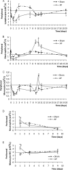
Severe sepsis is associated with early release of inflammatory mediators that contribute to the morbidity and mortality observed during the first stages of this syndrome. Although sepsis is a deadly, acute disease, high mortality rates have been observed in patients displaying evidence of sepsis-induced immune deactivation. Although the contribution of experimental models to the knowledge of pathophysiological and therapeutic aspects of human sepsis is undeniable, most of the current studies using animal models have focused on the acute, proinflammatory phase. We developed a murine model that reproduces the early acute phases but also the long-term consequences of human sepsis. We induced polymicrobial acute peritonitis (AP) by establishing a surgical connection between the cecum and the peritoneum, allowing the exit of intestinal bacteria. Using this model, we observed an acute phase with high mortality, leukopenia, increased interleukin-6 levels, bacteremia, and neutrophil activation. A peak of leukocytosis on day 9 or 10 revealed the persistence of the infection within the lung and liver, with inflammatory hepatic damage being shown by histological examination. Long-term (20 days) derangements in both innate and adaptive immune responses were found, as demonstrated by impaired systemic tumor necrosis factor alpha production in response to an inflammatory stimulus; a decreased primary humoral immune response and T cell proliferation, associated with an increased number of myeloid suppressor cells (Gr-1(+) CD11b(+)) in the spleen; and a low clearance capacity. This model provides a good approach to attempt novel therapeutic interventions directed to augmenting host immunity during late sepsis.
Figures


Similar articles
-
Increased mortality and altered immunity in neonatal sepsis produced by generalized peritonitis.Shock. 2007 Dec;28(6):675-683. doi: 10.1097/SHK.0b013e3180556d09. Shock. 2007. PMID: 17621256
-
Increased inflammation and lethality of Dusp1-/- mice in polymicrobial peritonitis models.Immunology. 2010 Nov;131(3):395-404. doi: 10.1111/j.1365-2567.2010.03313.x. Immunology. 2010. PMID: 20561086 Free PMC article.
-
Cecal ligation puncture procedure.J Vis Exp. 2011 May 7;(51):2860. doi: 10.3791/2860. J Vis Exp. 2011. PMID: 21587163 Free PMC article.
-
Immune dysfunction in murine polymicrobial sepsis: mediators, macrophages, lymphocytes and apoptosis.Shock. 1996;6 Suppl 1:S27-38. Shock. 1996. PMID: 8828095 Review.
-
Innate immune response to peritoneal bacterial infection.Int Rev Cell Mol Biol. 2022;371:43-61. doi: 10.1016/bs.ircmb.2022.04.014. Epub 2022 Jul 12. Int Rev Cell Mol Biol. 2022. PMID: 35965000 Review.
Cited by
-
Polymicrobial sepsis and non-specific immunization induce adaptive immunosuppression to a similar degree.PLoS One. 2018 Feb 7;13(2):e0192197. doi: 10.1371/journal.pone.0192197. eCollection 2018. PLoS One. 2018. PMID: 29415028 Free PMC article.
-
TRAIL induces neutrophil apoptosis and dampens sepsis-induced organ injury in murine colon ascendens stent peritonitis.PLoS One. 2014 Jun 2;9(6):e97451. doi: 10.1371/journal.pone.0097451. eCollection 2014. PLoS One. 2014. PMID: 24887152 Free PMC article.
-
Curcumol Targeting PAX8 Inhibits Ovarian Cancer Cell Migration and Invasion and Increases Chemotherapy Sensitivity of Niraparib.J Oncol. 2022 May 2;2022:3941630. doi: 10.1155/2022/3941630. eCollection 2022. J Oncol. 2022. PMID: 35548853 Free PMC article.
-
The microbial composition of the initial insult can predict the prognosis of experimental sepsis.Sci Rep. 2021 Nov 23;11(1):22772. doi: 10.1038/s41598-021-02129-x. Sci Rep. 2021. PMID: 34815465 Free PMC article.
-
Nicotinamide Mononucleotide Administration Triggers Macrophages Reprogramming and Alleviates Inflammation During Sepsis Induced by Experimental Peritonitis.Front Mol Biosci. 2022 Jun 27;9:895028. doi: 10.3389/fmolb.2022.895028. eCollection 2022. Front Mol Biosci. 2022. PMID: 35832733 Free PMC article.
References
-
- Adib-Conquy, M., and J. M. Cavaillon. 2009. Compensatory anti-inflammatory response syndrome. Thromb. Haemost. 101:36-47. - PubMed
-
- Aldridge, A. J. 2002. Role of the neutrophil in septic shock and the adult respiratory distress syndrome. Eur. J. Surg. 168:204-214. - PubMed
-
- Angus, D. C., and R. S. Wax. 2001. Epidemiology of sepsis: an update. Crit. Care Med. 29:S109-S116. - PubMed
-
- Atochina, O., T. Daly-Engel, D. Piskorska, E. McGuire, and D. A. Harn. 2001. A schistosome-expressed immunomodulatory glycoconjugate expands peritoneal Grl(+) macrophages that suppress naive CD4(+) T cell proliferation via an IFN-gamma and nitric oxide-dependent mechanism. J. Immunol. 167:4293-4302. - PubMed
-
- Bengualid, V., H. Singh, V. Singh, and J. Berger. 2008. An increase in the incidence of anaerobic bacteremia: true for tertiary care referral centers but not for community hospitals? Clin. Infect. Dis. 46:323-324. - PubMed
Publication types
MeSH terms
Substances
LinkOut - more resources
Full Text Sources
Other Literature Sources
Medical
Research Materials

