Comparative Study
. 2011 Jan 4;123(1):53-61.
doi: 10.1161/CIRCULATIONAHA.110.970673. Epub 2010 Dec 20.

Mitogen-activated protein kinase inhibitors improve heart function and prevent fibrosis in cardiomyopathy caused by mutation in lamin A/C gene

Affiliations
Comparative Study

Mitogen-activated protein kinase inhibitors improve heart function and prevent fibrosis in cardiomyopathy caused by mutation in lamin A/C gene

Wei Wu et al. Circulation. .

Abstract

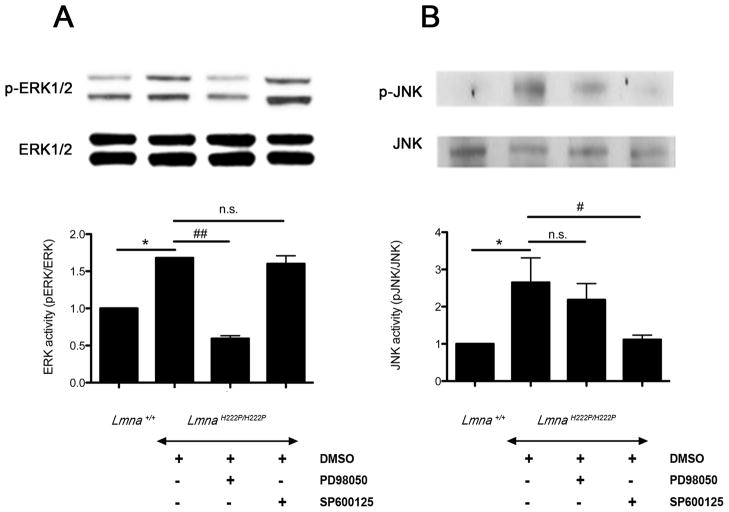
Background: Mutations in the lamin A/C gene, LMNA, can cause dilated cardiomyopathy. We have shown abnormal activation of the extracellular signal-regulated kinase (ERK) and the c-jun N-terminal kinase (JNK) branches of the mitogen-activated protein kinase signaling cascade in hearts from Lmna(H222P/H222P) mice that develop dilated cardiomyopathy. We recently showed that partial inhibition of ERK and JNK signaling before the onset of cardiomyopathy in Lmna(H222P/H222P) mice prevented the development of left ventricle dilatation and decreased cardiac ejection fraction at a time when they occurred in untreated mice.

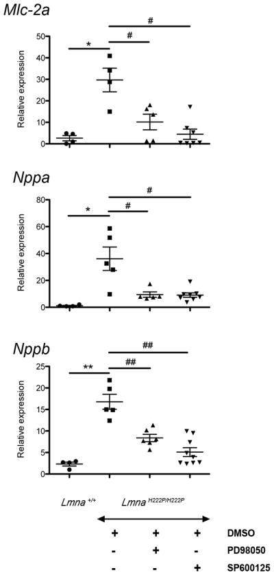
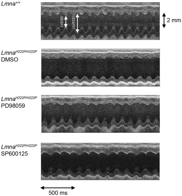
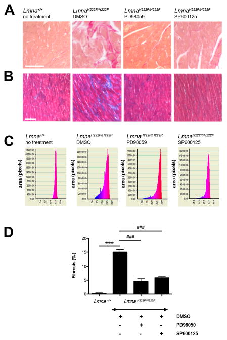
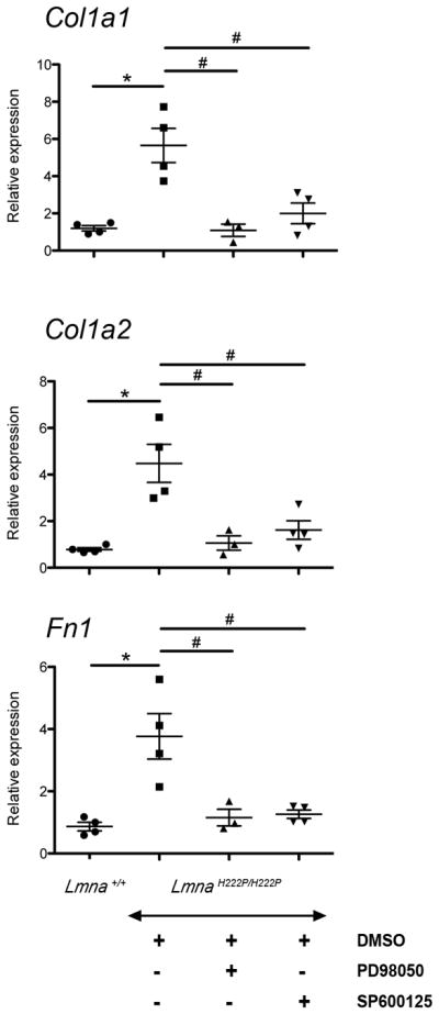
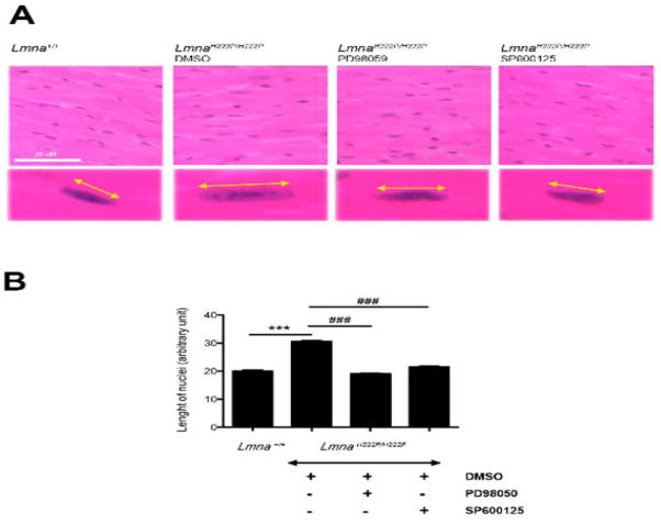
Methods and results: To determine whether pharmacological inhibitors of ERK and JNK signaling could be clinically useful to treat cardiomyopathy caused by LMNA mutation, we administered them to Lmna(H222P/H222P) mice after they developed left ventricular dilatation and decreased ejection fraction. Lmna(H222P/H222P) mice were treated with ERK and JNK signaling inhibitors from 16 to 20 or, in pilot experiments, 19 to 24 weeks of age. The inhibitors blocked increased expression of RNAs encoding natriuretic peptide precursors and proteins involved in sarcomere architecture that occurred in placebo-treated mice. Echocardiography and histological analysis demonstrated that treatment prevented left ventricular end-systolic dilatation, increased ejection fraction, and decreased myocardial fibrosis.

Conclusion: Inhibitors of ERK and JNK signaling could potentially be used to treat humans with cardiomyopathy caused by LMNA mutations.


Figures

Figure 1
Figure 1
(A) Representative immunoblots using antibodies against phophorylated ERK1/2 (p-ERK) and total ERK1/2 (ERK) and (B) against phophorylated JNK (p-JNK) and total JNK to probe proteins extracted from hearts from LmnaH222P/H222P mice treated with PD98059, SP600125 or DMSO. Blots of proteins extracted from hearts of Lmna+/+ mice are shown for comparison. The graphs show quantification of (A) pERK/total ERK and (B) pJNK/total JNK. n=4 in each group. Comparison between DMSO-treated LmnaH222P/H222P mice and Lmna+/+ mice; *P<0.05. Comparison between PD98059-treated and SP600125-treated and DMSO-treated LmnaH222P/H222P mice; #P<0.05, ##P<0.005, n.s.: not significant.
Figure 2
Figure 2
Effect of PD98059 and SP600125 on cardiac expression of natriuretic peptides and myosin light chain in LmnaH222P/H222P mice. Dot diagrams indicate the expression levels of Mlc-2a mRNA encoding the cardiac isoform of myosin light chain, Nppa mRNA encoding the atrial natriuretic factor and Nppb encoding the brain natriuretic peptide in hearts from Lmna+/+ mice and LmnaH222P/H222P mice treated with PD98059, SP600125 or DMSO. n=4 in each group. Values were obtained using the ΔΔCT method using Gapdh as housekeeping gene (see Full Materials and Methods). *P<0.05, **P<0.005, #P<0.05, ##P<0.005.
Figure 3
Figure 3
Representative transthoracic M-mode echocardiographic tracings from LmnaH222P/H222P mice treated with PD98059, SP600125 or DMSO. Tracings from Lmna+/+ mice are shown for comparison. LVESD and LVEDD are indicated.
Figure 4
Figure 4
(A) Sirius red and (B) Gomori’s trichrome staining of cross-sections of hearts from LmnaH222P/H222P mice treated with PD98059, SP600125 or DMSO. A cross-section of heart from a Lmna+/+ mouse is shown for comparison. Scale bar: 50 μm. (C) Quantification of fibrotic area in hearts from mice. n=3 in each group. Y-axis corresponds to the area (pixels) and X-axis represents the color spectrum (red corresponds to the muscle tissue and blue corresponds to the connective tissue). (D) Bars indicate the percentage of fibrosis per surface area of myocardium examined in hearts from Lmna+/+ mice and LmnaH222P/H222P mice treated with PD98059, SP600125 or DMSO. n=3 in each group. ***P<0.0005, ###P<0.0005.
Figure 5
Figure 5
Effect of PD98059 and SP600125 on cardiac expression of genes encoding collagen and fibronectin in LmnaH222P/H222P mice. Dot diagrams indicate the expression of Col1a1, Col1a2 and Fn1 in heart from Lmna+/+ mice and LmnaH222P/H222P mice treated with PD98059, SP600125 or DMSO. n=3 in each group. Values were obtained using the ΔΔCT method using Gapdh as housekeeping gene (see Full Materials and Methods). *P<0.05, #P<0.05.
Figure 6
Figure 6
(A) Histological analysis of cross-sections of hearts from LmnaH222P/H222P mice treated with PD98059, SP600125 or DMSO. Heart from a Lmna+/+ mouse is shown for comparison. Sections are stained with hematoxylin and eosin. Yellow lines with arrowheads demonstrate the measurement of nuclear length. Scale bar: 25 μm. (B) Quantification of nuclear elongation in cardiomyocytes from mice. Cardiomyocyte nuclei were measured along the yellow lines with arrowheads. Bars indicate the length of cardiomyocyte nuclei in the indicated hearts. Values are means ± SEM for n=150, 290, 690 and 575 cardiomyocytes from Lmna+/+ mice, DMSO-treated LmnaH222P/H222P mice, PD98059-treated LmnaH222P/H222P mice, and SP600125-treated LmnaH222P/H222P mice, respectively. ***P<0.0005, ###P<0.0005.

References

    1. Taylor MRG, Carniel E, Mestroni L. Cardiomyopathy, familial dilated. Orphanet J Rare Dis. 2006;13(1):27. - PMC - PubMed
    1. Worman HJ, Fong LG, Muchir A, Young SG. Laminopathies and the long strange trip from basic cell biology to therapy. J Clin Invest. 2009;119:1825–1836. - PMC - PubMed
    1. Bonne G, Di Barletta MR, Varnous S, Becane HM, Hammouda EH, Merlini L, Muntoni F, Greenberg CR, Gary F, Urtizberea JA, Duboc D, Fardeau M, Toniolo D, Schwartz K. Mutations in the gene encoding lamin A/C cause autosomal dominant Emery-Dreifuss muscular dystrophy. Nat Genet. 1999;21:285–288. - PubMed
    1. Muchir A, Bonne G, van der Kooi AJ, van Meegen M, Baas F, Bolhuis PA, de Visser M, Schwartz K. Identification of mutations in the gene encoding lamins A/C in autosomal dominant limb girdle muscular dystrophy with atrioventricular conduction disturbances (LGMD1B) Hum Mol Genet. 2000;9:1453–1459. - PubMed
    1. Fatkin D, MacRae C, Sasaki T, Wolff MR, Porcu M, Frenneaux M, Atherton J, Vidaillet HJ, Jr, Spudich S, De Girolami U, Seidman JG, Seidman C, Muntoni F, Müehle G, Johnson W, McDonough B. Missense mutations in the rod domain of the lamin A/C gene as causes of dilated cardiomyopathy and conduction-system disease. N Engl J Med. 1999;341:1715–1724. - PubMed

Publication types

MeSH terms