Review
. 2011 Mar-Apr;5(2):97-105.
doi: 10.4161/cam.5.2.14374. Epub 2011 Mar 1.

Non-clustered protocadherin

Affiliations
Review

Non-clustered protocadherin

Soo-Young Kim et al. Cell Adh Migr. 2011 Mar-Apr.

Abstract

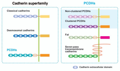
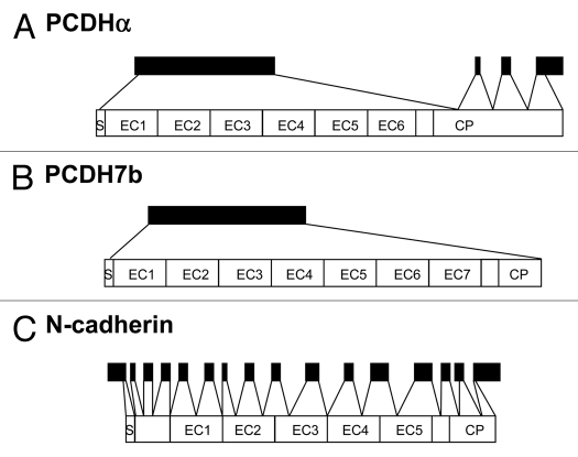
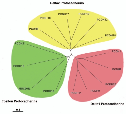
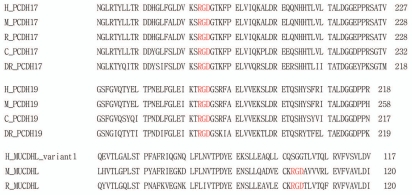
The cadherin family is classified into classical cadherins, desmosomal cadherins and protocadherins (PCDHs). Genomic structures distinguish between PCDHs and other cadherins, and between clustered and non-clustered PCDHs. The phylogenetic analysis with full sequences of non-clustered PCDHs enabled them to be further classified into three subgroups: δ1 (PCDH1, PCDH7, PCDH9, PCDH11 and PCDH20), δ2 (PCDH8, PCDH10, PCDH12, PCDH17, PCDH18 and PCDH19) and ε (PCDH15, PCDH16, PCDH21 and MUCDHL). ε-PCDH members except PCDH21 have either higher or lower numbers of cadherin repeats than those of other PCDHs. Non-clustered PCDHs are expressed predominantly in the nervous system and have spatiotemporally diverse expression patterns. Especially, the region-specific expressions of non-clustered PCDHs have been observed in cortical area of early postnatal stage and in caudate putaman and/or hippocampal formation of mature brains, suggesting that non-clustered PCDHs play roles in the circuit formation and maintenance. The non-clustered PCDHs appear to have homophilic/heterophilc cell-cell adhesion properties, and each member has diverse cell signaling partnership distinct from those of other members (PCDH7/TAF1; PCDH8/TAO2β; PCDH10/Nap1; PCDH11/β-catenin; PCDH18/mDab1). Furthermore, each PCDH has several isoforms with differential cytoplasmic sequences, suggesting that one PCDH isoform could activate intracellular signaling differential from other isoforms. These facts suggest that non-clustered PCDHs play roles as a mediator of a regulator of other molecules as well as cell-cell adhesion. Furthermore, some non-clustered PCDHs have been considered to be involved in neuronal diseases such as autism-spectrum disorders, schizophrenia, and female-limited epilepsy and cognitive impairment, suggesting that they play multiple, tightly regulated roles in normal brain function. In addition, some non-clustered PCDHs have been suggested as candidate tumor suppressor genes in several tissues. Although molecular adhesive and regulatory properties of some PCDHs began to be unveiled, the endeavor to understand the molecular mechanism of non-clustered PCDH is still in its infancy and requires future study.


Figures

Figure 1
Figure 1
Classification of cadherin superfamily including PCDHs. All cadherin superfamily members have calcium-binding cadherin repeats. The number of cadherin repeats varies from one subfamily to another. On average, a single cadherin repeat contains 110 amino acids in length. PCDHs are largely classified into the clustered PCDHs and non-clustered PCDHs, yet also include fats and seven-pass transmembrane cadherins in some cases. The clustered PCDHs are consisted of the PCDHα, β and γ family, which is clustered in a small genome locus. Non-clustered PCDHs are scattered in several genome loci.
Figure 2
Figure 2
Comparison of the genomic organization of α-PCDHs (A), mouse PCDH7b (B) and N-cadherin gene (C).
Figure 3
Figure 3
Phylogenetic tree of human non-clustered PCDHs on the basis of their total protein sequences. Multiple alignments and phylogenetic analysis were performed using the ClustalW2 program (www.ebi.ac.uk/Tools/clustalw2/), and the tree was visualized using the Treeview program (http://taxonomy.zoology.gla.ac.uk/rod/). The scale bar represents the substitution rate of amino acid per ten. All sequences used were obtained from the National Center for Biotechnology Information (NCBI). Accession numbers are as follows: PCDH1 (NM_002587), PCDH7 (NM_002589), PCDH8 (NM_002590), PCDH9 (NM_020403), PCDH10 (NM_032961), PCDH11 (NM_014522), PCDH12 (NM_016580), PCDH15 (NM_033056), PCDH16 (NM_003737), PCDH17 (NM_001040429), PCDH18 (NM_019035), PCDH19 (NM_020766), PCDH20 (NM_022843), PCDH21 (NM_033100) and MUCDHL (NM_021924).
Figure 4
Figure 4
The extracellular domain sequences of PCDH17, PCDH19 and MUCDHL. The RGD motif is found in the extracellular domains of PCDH17, PCDH19 and MUCDHL. The RGD motif is an essential residue for integrin-dependent cell adhesion activity. Other species are: H, human; M, mouse; R, rat; C, chick; DR, Danio rerio. PCDH17 (H, NM_014459; M, XM_127786; R, XM_224389; C, XM_417021; DR, XM_684743), PCDH19 (H, NM_001105243; M, NM_001105245; C, NM_001098607; DR, NP_001120991), MUCDHL (H, NM_021924; M, NM_028069; R, NM_138525).
Figure 5
Figure 5
Schematic overview of intracellular signaling proteins bound to the cytoplasmic domain of each PCDH. (A) PCDH7c (PCDH7c and 7c1, but not PCDH7a or 7b) has CM1, CM2 and CM3 motifs, and CM3 could be bound by PP1α. This interaction inactivates PP1α., In addition, all PCDH7 isoforms interact with histone-regulating protein TAF1. The PCDH7-TAF1 interaction is involved in retinal axon initiation and elongation in developing retinal ganglion cells. (B) All PCDH11Y isoforms (11Ya, Yb and Yc) have the binding sequences to δ-catenins, however, only PCDH11Yc (but not 11Ya or 11Yb) has CM1, CM2 and CM3 motifs. PCDH11Y is biochemically associated with δ-catenin, and this interaction might affect wnt signaling and tumorgenesis. (C) PCDH8/arcadlin binds to a serine-threonine kinase TAO2δ. This interaction causes N-cadherin endocytosis at synaptic membrane in a p38 MAPK-dependent manner. (D) PCDH10/OL-PC interacts with Nap1, and this interaction recruits WAVE1, a Nap1 binding protein, to cell-cell contact sites. The formation of PCDH10/Nap1/WAVE complex regulates actin assembly, and subsequently promotes the migration of cells. (E) PCDH18 is associated with mDab1, and this interaction might be involved in the correct formation of cortical nerve cell layers.

Similar articles

Cited by

References

    1. Nollet F, Kools P, van Roy F. Phylogenetic analysis of the cadherin superfamily allows identification of six major subfamilies besides several solitary members. J Mol Biol. 2000;299:551–572. - PubMed
    1. Halbleib JM, Nelson WJ. Cadherins in development: cell adhesion, sorting and tissue morphogenesis. Genes Dev. 2006;20:3199–3214. - PubMed
    1. Morishita H, Yagi T. Protocadherin family: diversity, structure and function. Curr Opin Cell Biol. 2007;19:584–592. - PubMed
    1. Wu Q, Maniatis T. A striking organization of a large family of human neural cadherin-like cell adhesion genes. Cell. 1999;97:779–790. - PubMed
    1. Redies C, Vanhalst K, Roy F. delta-Protocadherins: unique structures and functions. Cell Mol Life Sci. 2005;62:2840–2852. - PMC - PubMed

Publication types

MeSH terms

LinkOut - more resources