

Comment
. 2011 Jan;13(1):3-5; author reply 5-7.
doi: 10.1038/ncb0111-3.
Reducing background fluorescence reveals adhesions in 3D matrices
- PMID: 21173800
- PMCID: PMC3083631
- DOI: 10.1038/ncb0111-3
Item in Clipboard
Comment
Reducing background fluorescence reveals adhesions in 3D matrices
Nat Cell Biol.
2011 Jan.
Erratum in
- Nat Cell Biol. 2012 Dec;14(12):1344
No abstract available
Figures
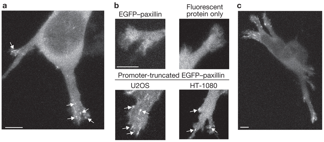
Cell-matrix adhesions are detected in 3D collagen gels. (a) Z-projection of a multi-polar U2OS cell expressing EGFP–paxillin under the control of a truncated promoter, with adhesions on the distal sections of the protrusions (arrows). See Supplementary Information, Fig. S1a–f for more images. (b) Z-projection images of protrusions from cells expressing the indicated construct. The cell expressing a low level of normal EGFP–paxillin (upper left) has a diffuse cytoplasmic fluorescence, similar in appearance to the cell expressing the fluorescent protein TagRFP-T alone (upper-right). Adhesions (arrows) are detectable in U2OS cells (bottom left) and HT-1080 cells (bottom right) transfected with a plasmid encoding promoter-truncated EGFP–paxillin, which expresses at a lower level than EGFP–paxillin and decreases background cytoplasmic fluorescence. (c) Z-projection of a U2OS cell expressing promoter-truncated EGFP–vinculin. Scale bars, 5 µm.
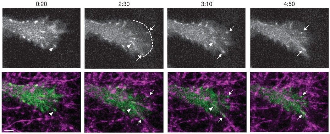
Dynamics of cell-matrix adhesions in 3D culture. Frames are taken from Supplementary Information, Video S1. The top row shows Z-projections of a protrusion end from a U2OS cell that was transfected with a plasmid encoding promoter-truncated EGFP–paxillin. The bottom row shows the image from the top row (green) overlayed with a reflectance image of the collagen fibres (magenta). Time indicates the min:s since the beginning of the movie. At 0:20, a small adhesion (arrowhead) moves rearward, pulling a collagen fibre. At 2:30, a new protrusion (boundary shown by dotted line) pauses, two new adhesions form at the leading edge (arrows), and the earlier adhesion (arrowhead) has elongated while travelling rearward. At 3:10, the two new adhesions (arrows) continue to grow and translocate with the attached collagen fibres. By 4:50, the new adhesions have continued to grow and move rearward while the early adhesion (arrowhead in previous panels) is no longer visible. Scale bar,2 µm
Comment on
-
A distinctive role for focal adhesion proteins in three-dimensional cell motility.Nat Cell Biol. 2010 Jun;12(6):598-604. doi: 10.1038/ncb2062. Epub 2010 May 16. Nat Cell Biol. 2010. PMID: 20473295 Free PMC article.
References
Publication types
MeSH terms
Substances
Grants and funding
LinkOut - more resources
Full Text Sources
Other Literature Sources
