Review
. 2011 Jan;248(1):61-74.
doi: 10.1007/s00709-010-0252-3. Epub 2010 Dec 21.

Plasmodesmata during development: re-examination of the importance of primary, secondary, and branched plasmodesmata structure versus function

Affiliations
Review

Plasmodesmata during development: re-examination of the importance of primary, secondary, and branched plasmodesmata structure versus function

Tessa M Burch-Smith et al. Protoplasma. 2011 Jan.

Abstract

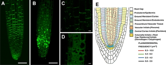
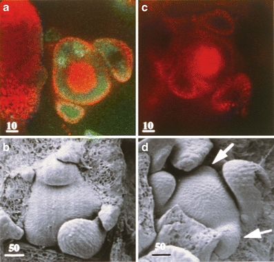
Plasmodesmata (PD) structure and function vary temporally and spatially during all stages of plant development. PD that originate during, or post, cell division are designated as primary or secondary according to classical terminology. PD structure may be simple, twinned, or branched. Studies of PD during leaf, root, and embryo development have lead to the generalization that cells in less mature tissues contain predominantly simple PD. New quantitative analyses reveal that twinned and branched PD also occur in immature tissues. New data also highlight the versatility of viral movement proteins as tags for labeling PD in immature tissues as well as PD in mature tissues. A summary of the formation and function of primary, secondary, and branched PD during leaf, trichome, embryo, apical meristem, vascular cambium, and root development underscores the remarkable and indispensible plant-specific intercellular communication system that is mediated by PD.


Figures

Fig. 1
Fig. 1
PD structures and how they form. a, b Present different intermediate PD structures that may form during the creation of twinned PD by fission of a simple PD. a Initiation of PD doubling at one site that results in Y- and V-shaped intermediates. b Initiation of PD doubling at two sites that results in X- and H-shaped intermediates. c How an H-shaped PD may become further branched and ultimately contain a central cavity (c). d A mechanistic model for how twinned PD or branched PD arise that derives from the laying down of new cell wall material (dark grey shading) as the cell wall (light grey shading) expands laterally. New cell wall material may initiate the branching of PD and then ultimately separate the two newly formed PD channels. While the cell wall is not drawn in panels a, b, and c, cell wall expansion likely also drives the formation of the Y-, V-, X-, and H-shaped PD, twinned PD, and multiple-branched PD. Note the PD channels diagrammed in all panels represent the cell membrane bound channel without the central ER-derived desmotubule. The model drawn in D is a simplification of a similar model in Ehlers and van Bel (2010)
Fig. 2
Fig. 2
Localization of viral movement proteins in Arabidopsis seedling root tips. TMV-MP-GFP constitutively expressed from the CaMV minimal 35S promoter in a 5-day-old transgenic Arabidopsis seedling root tips localizes predominantly to plasmodesmata in transverse cell walls (a) containing high densities of primary PD while PLRV MP17-GFP expressed from the same promoter fails to localize to plasmodesmata in root tips (b). Images are projections of confocal stacks; scale bar represents 50 μm. Higher magnification confocal images of TMV-MP-GFP (c) and PLRV MP17-GFP expression (d) in root tip epidermal cells reveal that TMV-MP-GFP is localized largely to punctae in transverse cell walls while PLRV MP17-GFP is retained in the cytoplasm and forms cytoplasmic aggregates. Scale bar represents 5 μm. e A graphic representation of the distribution and frequency of predominantly primary plasmodesmata in 1-week-old Arabidopsis root tips demonstrates that transverse cell walls in all cell layers of the root tip contain a higher density of plasmodesmata than longitudinal cell walls. e Adapted from Fig. 4 of Zhu et al. and used with kind permission from Springer: Protoplasma
Fig. 3
Fig. 3
Movement of symplastic tracers during the transition to floral development in Arabidopsis. Arabidopsis grown under short-day (SD) conditions were loaded through their leaf petioles with 8-hydroxypyrene 1,3,6,-trisulfonic acid (HPTS), molecular mass 520 Da. Following 39 and 42 SDs, seedlings have 15 (a, b) and 19 leaves (c, d). a At 39 SD, plants are still vegetative and unload HPTS into their apices following transport via the vascular system from leaf petioles. The image is a confocal optical section and documents that HPTS does not unload in the center of the meristem (Gisel et al. 1999); however, HPTS does unload throughout leaf primordia seen around the circumference of the meristem. c At 42 SD, plants have initiated reproductive development and no longer unload HPTS into their apices. Vegetative versus reproductive apices are distinguished by the types of primordia they produce; leaf primordia are pointy versus floral primordia have a rounded shape (arrows in (d)). b Scanning electron micrograph (SEM) of a 39-SD-old apex reveals leaf primordia around the SAM. d SEM of a 42-SD apex reveals leaf primordia as well as newly arising floral primordia. Data were obtained as described in Gisel et al.

References

    1. Baum SF, Rost TL. Root apical organization in Arabidops thaliana 1. Root cap and protoderm. Protoplasma. 1996;192:178–188. doi: 10.1007/BF01273890. - DOI
    1. Benfey PN, Scheres B. Root development. Curr Biol. 2000;10:R813–R815. doi: 10.1016/S0960-9822(00)00814-9. - DOI - PubMed
    1. Benitez-Alfonso Y, Cilia M, San Roman A, Thomas C, Maule A, Hearn S, Jackson D. Control of Arabidopsis meristem development by thioredoxin-dependent regulation of intercellular transport. Proc Natl Acad Sci USA. 2009;106:3615–3620. doi: 10.1073/pnas.0808717106. - DOI - PMC - PubMed
    1. Benitez-Alfonso Y, Jackson D, Maule A (2010). Redox regulation of intercellular transport. Protoplasma (in press). - PubMed
    1. Brady SM, Orlando DA, Lee JY, Wang JY, Koch J, Dinneny JR, Mace D, Ohler U, Benfey PN. A high-resolution root spatiotemporal map reveals dominant expression patterns. Science. 2007;318:801–806. doi: 10.1126/science.1146265. - DOI - PubMed

Publication types

MeSH terms