

Non-fluent aphasia and neural reorganization after speech therapy: insights from human sleep electrophysiology and functional magnetic resonance imaging
- PMID: 21175013
- PMCID: PMC3058764
Non-fluent aphasia and neural reorganization after speech therapy: insights from human sleep electrophysiology and functional magnetic resonance imaging
Abstract
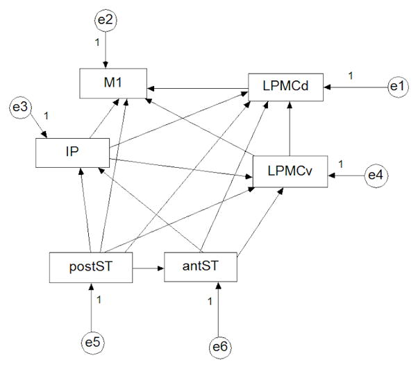
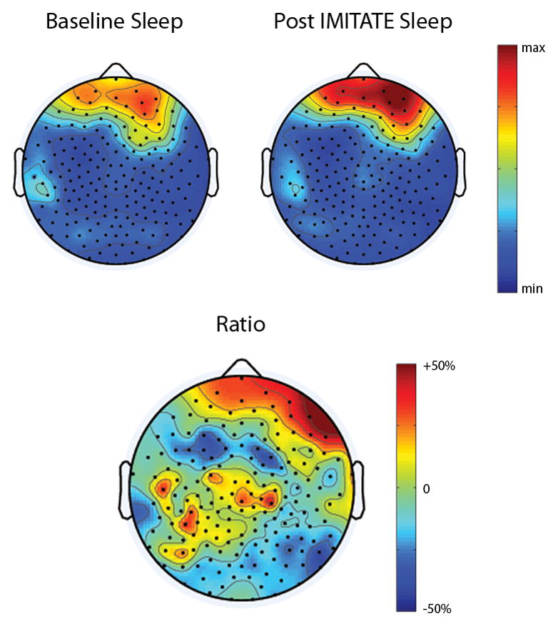
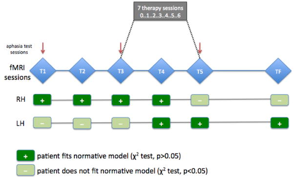
Stroke is associated with long-term functional deficits. Behavioral interventions are often effective in promoting functional recovery and plastic changes. Recent studies in normal subjects have shown that sleep, and particularly slow wave activity (SWA), is tied to local brain plasticity and may be used as a sensitive marker of local cortical reorganization after stroke. In a pilot study, we assessed the local changes induced by a single exposure to a therapeutic session of IMITATE (Intensive Mouth Imitation and Talking for Aphasia Therapeutic Effects), a behavioral therapy used for recovery in patients with post-stroke aphasia. In addition, we measured brain activity changes with functional magnetic resonance imaging (fMRI) in a language observation task before, during and after the full IMITATE rehabilitative program. Speech production improved both after a single exposure and the full therapy program as measured by the Western Aphasia Battery (WAB) Repetition subscale. We found that IMITATE induced reorganization in functionally-connected, speech-relevant areas in the left hemisphere. These preliminary results suggest that sleep hd-EEGs, and the topographical analysis of SWA parameters, are well suited to investigate brain plastic changes underpinning functional recovery in neurological disorders.
Figures
Similar articles
-
Plastic changes following imitation-based speech and language therapy for aphasia: a high-density sleep EEG study.Neurorehabil Neural Repair. 2014 Feb;28(2):129-38. doi: 10.1177/1545968313498651. Epub 2013 Aug 26. Neurorehabil Neural Repair. 2014. PMID: 23980019
-
Effects of low-frequency repetitive transcranial magnetic stimulation combined with intensive speech therapy on cerebral blood flow in post-stroke aphasia.Transl Stroke Res. 2015 Oct;6(5):365-74. doi: 10.1007/s12975-015-0417-7. Epub 2015 Aug 7. Transl Stroke Res. 2015. PMID: 26245774
-
Anterior temporal lobe connectivity correlates with functional outcome after aphasic stroke.Brain. 2009 Dec;132(Pt 12):3428-42. doi: 10.1093/brain/awp270. Brain. 2009. PMID: 19903736 Free PMC article.
-
Functional MRI of language in aphasia: a review of the literature and the methodological challenges.Neuropsychol Rev. 2007 Jun;17(2):157-77. doi: 10.1007/s11065-007-9024-z. Epub 2007 May 25. Neuropsychol Rev. 2007. PMID: 17525865 Free PMC article. Review.
-
[Functional neuroimaging and the treatment of aphasia: speech therapy and repetitive transcranial magnetic stimulation].Rev Neurol (Paris). 2008 May;164 Suppl 3:S45-8. doi: 10.1016/S0035-3787(08)73290-9. Rev Neurol (Paris). 2008. PMID: 18675046 Review. French.
Cited by
-
Non-invasive Brain Stimulation in the Treatment of Post-stroke and Neurodegenerative Aphasia: Parallels, Differences, and Lessons Learned.Front Hum Neurosci. 2017 Jan 23;10:675. doi: 10.3389/fnhum.2016.00675. eCollection 2016. Front Hum Neurosci. 2017. PMID: 28167904 Free PMC article. Review.
-
Changes in task-based effective connectivity in language networks following rehabilitation in post-stroke patients with aphasia.Front Hum Neurosci. 2015 Jun 9;9:316. doi: 10.3389/fnhum.2015.00316. eCollection 2015. Front Hum Neurosci. 2015. PMID: 26106314 Free PMC article.
-
Neuroimaging in aphasia treatment research: standards for establishing the effects of treatment.Neuroimage. 2013 Aug 1;76:428-35. doi: 10.1016/j.neuroimage.2012.10.011. Epub 2012 Oct 9. Neuroimage. 2013. PMID: 23063559 Free PMC article. Review.
-
Brain repair after stroke--a novel neurological model.Nat Rev Neurol. 2013 Dec;9(12):698-707. doi: 10.1038/nrneurol.2013.222. Epub 2013 Nov 12. Nat Rev Neurol. 2013. PMID: 24217509 Free PMC article. Review.
-
Tracking reorganization of large-scale effective connectivity in aphasia following right hemisphere stroke.Brain Lang. 2017 Jul;170:12-17. doi: 10.1016/j.bandl.2017.03.003. Epub 2017 Mar 29. Brain Lang. 2017. PMID: 28364641 Free PMC article.
References
-
- Buccino G, Binkofski F, Fink GR, Fadiga L, Fogassi L, Gallese V, Seitz RJ, Zilles K, Rizzolatti G, Freund HJ. Action observation activates premotor and parietal areas in a somatotopic manner: an fMRI study. Eur J Neurosci. 2001;13:400–404. - PubMed
-
- Buccino G, Vogt S, Ritzl A, Fink GR, Zilles K, Freund HJ, Rizzolatti G. Neural circuits underlying imitation learning of hand actions: An event-related fMRI study. Neuron. 2004;42:323–334. - PubMed
-
- Carmichael ST, Archibeque I, Luke L, Nolan T, Momiy J, Li S. Growth-associated gene expression after stroke: evidence for a growth-promoting region in peri-infarct cortex. Exp Neurol. 2005;193:291–311. - PubMed
Publication types
MeSH terms
Substances
Grants and funding
LinkOut - more resources
Full Text Sources
Medical