

doi: 10.1021/jm101359c.
Epub 2010 Dec 22.
Interactions of monoamine oxidases with the antiepileptic drug zonisamide: specificity of inhibition and structure of the human monoamine oxidase B complex
Affiliations
- PMID: 21175212
- PMCID: PMC3071873
- DOI: 10.1021/jm101359c
Item in Clipboard
Interactions of monoamine oxidases with the antiepileptic drug zonisamide: specificity of inhibition and structure of the human monoamine oxidase B complex
J Med Chem.
.
Abstract
The binding of zonisamide to purified, recombinant monoamine oxidases (MAOs) has been investigated. It is a competitive inhibitor of human MAO B (K(i) = 3.1 ± 0.3 μM), of rat MAO B (K(i) = 2.9 ± 0.5 μM), and of zebrafish MAO (K(i) = 30.8 ± 5.3 μM). No inhibition is observed with purified human or rat MAO A. The 1.8 Å structure of the MAO B complex demonstrates that it binds within the substrate cavity.
Figures
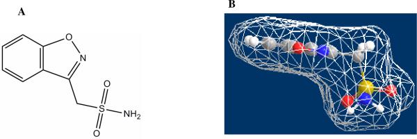
Structure of Zonisamide (A). Panel B shows the Chem3D representation of the zonisamide structure with a Connolly surface included after energy minimization. Carbons are in gray, hydrogens in white, nitrogens in blue, oxygens in red, and sulfur in yellow.
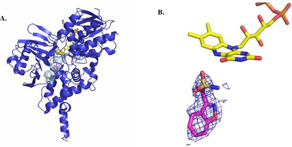
Structural properties of Zonisamide binding to human MAO B. (A) Ribbon diagram of the overall structure of human MAO B in complex with zonisamide. The substrate-binding cavity is represented as semi-transparent surface. Zonisamide is shown in magenta, FAD in yellow, and the MAO B chain trace in blue. (B) Weighted 2Fo-Fc electron density of bound zonisamide in the active site of MAO B. The map was calculated before inclusion in the model of the inhibitor and, therefore, it is fully unbiased. Zonisamide carbons are in magenta, flavin carbons in yellow, oxygens in red, nitrogens in blue, sulphurs in light brown, and phosphorous in orange. This figure as well as Figures 3 and 4 were produced with PyMol (www.pymol.org ).
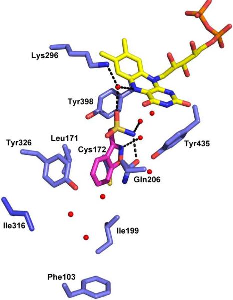
Structure of zonisamide in the active site of MAO B. Zonisamide carbons are in magenta, flavin carbons in yellow, protein carbons in light blue, oxygens in red, nitrogens in blue, sulphurs in light brown, and phosphorous in orange. Water molecules apparent in the electron density are depicted as red spheres. H-bonds suggested from distance and geometry considerations are denoted by dashed lines.
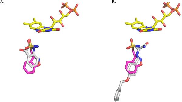
Superposition of zonisamide-MAO B complex with those with (A) isatin (PDB 2BK5) and (B) safinamide (PDB 2V5Z). The colors of residues and atoms are as in Figure 3. The carbon skeletons of isatin and safinamide structures are depicted in gray.
Similar articles
-
Leflunomide, a Reversible Monoamine Oxidase Inhibitor.Cent Nerv Syst Agents Med Chem. 2016;16(2):112-9. doi: 10.2174/1871524915666150824154329. Cent Nerv Syst Agents Med Chem. 2016. PMID: 26299850
-
Inhibition of monoamine oxidase by indole-5,6-dicarbonitrile derivatives.Bioorg Med Chem Lett. 2015 Mar 15;25(6):1206-11. doi: 10.1016/j.bmcl.2015.01.061. Epub 2015 Feb 4. Bioorg Med Chem Lett. 2015. PMID: 25701250
-
Three-dimensional structure of human monoamine oxidase A (MAO A): relation to the structures of rat MAO A and human MAO B.Proc Natl Acad Sci U S A. 2005 Sep 6;102(36):12684-9. doi: 10.1073/pnas.0505975102. Epub 2005 Aug 29. Proc Natl Acad Sci U S A. 2005. PMID: 16129825 Free PMC article.
-
Inhibitor design for monoamine oxidases.Curr Pharm Des. 2013;19(14):2529-39. doi: 10.2174/1381612811319140004. Curr Pharm Des. 2013. PMID: 23116392 Review.
-
[Computer modelling of monoaminoxidases].Biomed Khim. 2015 Mar-Apr;61(2):265-71. doi: 10.18097/PBMC20156102265. Biomed Khim. 2015. PMID: 25978392 Review. Russian.
Cited by
-
Coumarin-Chalcone Hybrids as Inhibitors of MAO-B: Biological Activity and In Silico Studies.Molecules. 2021 Apr 22;26(9):2430. doi: 10.3390/molecules26092430. Molecules. 2021. PMID: 33921982 Free PMC article.
-
Design, synthesis and evaluation of 1-(1,5-bis(4-substituted phenyl)-2-methyl-1H-pyrrol-3-yl)-N-methylmethanamines as SERT inhibitors with potential antidepressant action.RSC Med Chem. 2022 Oct 29;14(2):393-402. doi: 10.1039/d2md00243d. eCollection 2023 Feb 22. RSC Med Chem. 2022. PMID: 36846366 Free PMC article.
-
Zonisamide Enhances Motor Effects of Levodopa, Not of Apomorphine, in a Rat Model of Parkinson's Disease.Parkinsons Dis. 2018 Dec 18;2018:8626783. doi: 10.1155/2018/8626783. eCollection 2018. Parkinsons Dis. 2018. PMID: 30662707 Free PMC article.
-
Monoaminergic Mechanisms in Epilepsy May Offer Innovative Therapeutic Opportunity for Monoaminergic Multi-Target Drugs.Front Neurosci. 2016 Nov 10;10:492. doi: 10.3389/fnins.2016.00492. eCollection 2016. Front Neurosci. 2016. PMID: 27891070 Free PMC article. Review.
-
Computational Drug Target Screening through Protein Interaction Profiles.Sci Rep. 2016 Nov 15;6:36969. doi: 10.1038/srep36969. Sci Rep. 2016. PMID: 27845365 Free PMC article.
References
-
- Uno H, Kuokawa M, Masuda Y, Nishimara H. Studies on 3- substituted 1,2-benzisoxazole derivatives, 6. Synthesis of 3-(sulfamoylmethyl)-1,2-benzisoxazole derivatives and their anticonvulsant activities. J. Med. Chem. 1979;22:180–183. - PubMed
-
- Murata M. Novel Therapeutic Effects of the Anti-convulsant, Zonisamide, on Parkinson's Disease. Curr. Pharm. Des. 2004;10:687–693. - PubMed
-
- Yang LPH, Perry CM. Zonisamide in Parkinson's Disease. CNS Drugs. 2009;23:703–711. - PubMed
-
- Okada M, Kaneko S, Hirano T, Mizuno K, Kondo , Otani K, Fukushima Y. Effects of Zonisamide on Dopamminergic System. Epilepsy Res. 1995;22:193–205. - PubMed
Publication types
MeSH terms
Substances
Associated data
- Actions
Grants and funding
LinkOut - more resources
Full Text Sources
Chemical Information
