. 2011 Jan;6(1):53-66.
doi: 10.2217/rme.10.91.

Efficient generation and cryopreservation of cardiomyocytes derived from human embryonic stem cells

Affiliations

Efficient generation and cryopreservation of cardiomyocytes derived from human embryonic stem cells

Chunhui Xu et al. Regen Med. 2011 Jan.

Abstract

Aim: Human embryonic stem cells (hESCs) represent a novel cell source to treat diseases such as heart failure and for use in drug screening. In this study, we aim to promote efficient generation of cardiomyocytes from hESCs by combining the current optimal techniques of controlled growth of undifferentiated cells and specific induction for cardiac differentiation. We also aim to examine whether these methods are scalable and whether the differentiated cells can be cryopreserved.

Methods & results: hESCs were maintained without conditioned medium or feeders and were sequentially treated with activin A and bone morphogenetic protein-4 in a serum-free medium. This led to differentiation into cell populations containing high percentages of cardiomyocytes. The differentiated cells expressed appropriate cardiomyocyte markers and maintained contractility in culture, and the majority of the cells displayed working chamber (atrial and ventricular) type electrophysiological properties. In addition, the cell growth and differentiation process was adaptable to large culture formats. Moreover, the cardiomyocytes survived following cryopreservation, and viable cardiac grafts were detected after transplantation of cryopreserved cells into rat hearts following myocardial infarctions.

Conclusion: These results demonstrate that cardiomyocytes of high quality can be efficiently generated and cryopreserved using hESCs maintained in serum-free medium, a step forward towards the application of these cells to human clinical use or drug discovery.


Figures
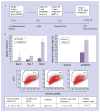
Figure 1
Figure 1. Cardiomyocyte differentiation induced by growth factor treatment in a serum-free medium
(A) Undifferentiated hESCs maintained in either MEF-CM or serum-free medium/GFs (stage 1) were treated sequentially with activin A (stage 2A) and BMP-4 (stage 2B) in RPMI/B27, followed by removal of growth factors and maintenance in RPMI/B27 (stage 3). Cells were analyzed for cardiomyocyte differentiation by ICC or flow cytometric analysis after stage 3. (B) Timing of cardiomyocyte generation. Confluent undifferentiated cells were induced to differentiate using the sequential treatment with activin A and BMP-4 method (A100/B10) or maintained in RPMI/B27 medium without growth factors (control); cultures were fixed at days 6, 7, 8 or 23 and analyzed for the presence of Nkx2.5-positive cells and for cells that were positive for both Nkx2.5 and α-actinin. Data are represented as mean ± standard deviation of three independent wells. (C) An example of differentiation of hESCs maintained in serum-free medium/GFs in small culture formats. Undifferentiated cells were seeded into six-well plates at 1 × 10⁵ cells/cm2 and induced to differentiate. Numerous beating cells were observed in these cultures at stage 3. Cells were harvested and analyzed for the percentage of cells expressing cTnI. The cell viability, determined by ethidium monoazide staining, and cell yields are presented as the mean ± standard deviation from triplicate wells. BMP: Bone morphogenetic protein; cTnI: Cardiac troponin I; D: Day; GF: Growth factor; hESC: Human embryonic stem cell; ICC: Immunocytochemical analysis; MEF-CM: Conditioned medium from mouse embryonic fibroblasts; Non-CM/GFs: Serum-free medium supplemented with growth factors.
Figure 2
Figure 2. Differentiation of human embryonic stem cells maintained in serum-free medium/GFs in large culture formats
Undifferentiated cells from independent cultures maintained in serum-free medium/GFs were seeded into T225 flasks at 1 × 10⁵ cells/cm2; differentiation was induced with the three stage growth factor-directed differentiation method. Similar to cultures in small formats, numerous beating cells were observed at stage three. Cells were harvested and analyzed for expression of cardiomyocyte-associated proteins, such as Nkx2.5 or α-actinin (indicated by the scatter plots). Cell viability was determined by ethidium monoazide staining. Yields of total cells and viable cardiac cells per flask as well as input undifferentiated hESC:viable cardiac cell ratio in each experiment are presented. hESC: Human embryonic stem cell.
Figure 3
Figure 3. Immunocytochemical analysis of cardiomyocytes derived from human embryonic stem cells using the growth factor differentiation method
At the end of differentiation stage 3, cells were dissociated, replated and cultured for an additional 6 days before immunocytochemical analysis. Nuclei were counterstained with 4′,6-diamidino-2-phenylindole (DAPI; blue). (A) Nkx2.5 (red), tropomyosin (green); (B) Nkx2.5 (red), cardiac troponin I (green); (C) Nkx2.5 (red), cardiac troponin T (green); (D) N-cadherin (red), sarcomeric myosin heavy chain (green); (E) Nkx2.5 (red), α-actinin (green); (F) cardiac troponin I (red), connexin 43 (green); (G) Enlarged image of (E) shows formation of striations in these cells. (H) Enlarged image of (F); (I) α-actinin (green); (J) Ki-67 (red); (K) merge of images (I & J) and DAPI; (L) Nkx2.5 (red); (M) Ki-67 (green); and (N) merge of images (L & M) and DAPI. Scale bar = 50 μm.
Figure 4
Figure 4. Electrophysiological analysis of cardiomyocytes derived from human embryonic stem cells using the growth factor differentiation method
Perforated-patch current-clamp recordings from two representative cardiomyocytes show spontaneous action potentials with distinct nodal-type (upper) and working chamber-type (lower) waveforms and characteristics.
Figure 5
Figure 5. Cryopreservation of cardiomyocytes derived from human embryonic stem cells
Cultures containing contracting cardiomyocytes were dissociated and cryopreserved using a controlled rate freezer. Cells before and after thawing were analyzed for cell viability, percentages of cardiac cells and cell recovery rates. (A) An example of flow cytometry analysis of samples before and after thawing. (B) Large-scale cryopreservation of human embryonic stem cell-derived cardiomyocytes. Differentiated cultures (containing 59% Nkx2.5+ cells; 86% viability) derived from human embryonic stem cells were cryopreserved at 1 × 10⁷ cells in 0.25 ml/vial, 4 × 10⁷ cells in 1.5 ml/vial and 8 × 10⁷ cells in 1.5 ml/vial. Triplicate vials were thawed and analyzed for percentages of cardiac cells, viability and recovery. Data are presented as mean ± standard deviation from triplicate vials. EMA: Ethidium monoazide.
Figure 6
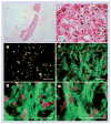
Figure 6. Immunohistochemical analysis of human cell grafts in the myocardium of infarcted rat hearts in the 1-week study
Freshly isolated cardiomyocytes or cryopreserved cardiomyocytes were resuspended in medium containing a prosurvival cocktail [10] and injected into the myocardium of rats that had received an ischemia–reperfusion injury. Hearts were harvested and processed for histological and immunohistochemical analysis 1 week after transplantation. As indicated by positive labeling for human nuclear antigen (HNA; brown diaminobenzidene reaction product in (A–D) and red immunofluorescence in (E & F)), human cell grafts were identified in both the group of animals that received freshly isolated cells (A,C & E) and in the group that received cryopreserved cells (B,D & F). Lower magnification images illustrate that graft sizes in the group that received freshly isolated cells (A) were comparable to those in the group that received cryopreserved cells (B). Double labeling of HNA with β-myosin heavy chain (red chromogen in (A–D)) indicated that the majority of the HNA-positive cells were cardiomyocytes, which was also confirmed by HNA/Nkx2.5 double labeling in (E & F) (red for HNA and green for Nkx2.5; cells labeled by both antibodies appear yellow). Scale bar = 500 μm for images in (A & B); scale bar = 50 μm for images in (C–F).
Figure 7
Figure 7. Immunohistochemical analysis of human cell grafts in the myocardium of infarcted rat hearts in the 4-week study
Cryopreserved cardiomyocytes were resuspended in medium containing a prosurvival cocktail [10] and injected into the myocardium of rats that had received an ischemia–reperfusion injury. Hearts were harvested and processed for histological and immunohistochemical analysis 4 weeks after transplantation. As indicated by positive labeling for human nuclear antigen (HNA; brown diaminobenzidene reaction product in (A & B) and red immunofluorescence in (C–F)), human cell grafts were identified in all animals as shown in a representative lower magnification image (A). Double labeling of HNA with β-myosin heavy chain (red chromogen in (A & B)) indicated that the majority of the HNA-positive cells were cardiomyocytes, which was also confirmed by HNA/Nkx2.5 double labeling in (C) (red for HNA and green for Nkx2.5; cells labeled by both antibodies appear yellow) and HNA/cardiac troponin I double labeling in (D–F) (red for HNA and green for cardiac troponin I). Higher magnifications of the image in (D) revealed the formation of muscle striations in (E & F). Scale bar = 500 μm for images in (A); scale bar = 50 μm for images in (B–F).

References

    1. Thomson JA, Itskovitz-Eldor J, Shapiro SS, et al. Embryonic stem cell lines derived from human blastocysts. Science. 1998;282:1145–1147. - PubMed
    1. Reubinoff BE, Pera MF, Fong CY, et al. Embryonic stem cell lines from human blastocysts: somatic differentiation in vitro. Nat Biotechnol. 2000;18(4):399–404. - PubMed
    1. Xue T, Cho HC, Akar FG, et al. Functional integration of electrically active cardiac derivatives from genetically engineered human embryonic stem cells with quiescent recipient ventricular cardiomyocytes: insights into the development of cell-based pacemakers. Circulation. 2005;111(1):11–20. - PubMed
    1. He JQ, Ma Y, Lee Y, et al. Human embryonic stem cells develop into multiple types of cardiac myocytes: action potential characterization. Circ Res. 2003;93(1):32–39. - PubMed
    1. Reppel M, Boettinger C, Hescheler J. β-adrenergic and muscarinic modulation of human embryonic stem cell-derived cardiomyocytes. Cell Physiol Biochem. 2004;14(4–6):187–196. - PubMed

Publication types

Substances