. 2011 Jan;175(1):97-112.
doi: 10.1667/RR2332.1.

Base damage immediately upstream from double-strand break ends is a more severe impediment to nonhomologous end joining than blocked 3'-termini

Affiliations

Base damage immediately upstream from double-strand break ends is a more severe impediment to nonhomologous end joining than blocked 3'-termini

Kamal Datta et al. Radiat Res. 2011 Jan.

Abstract

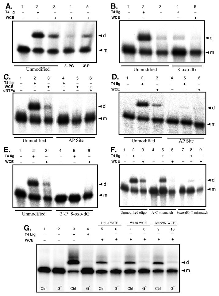
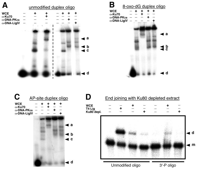
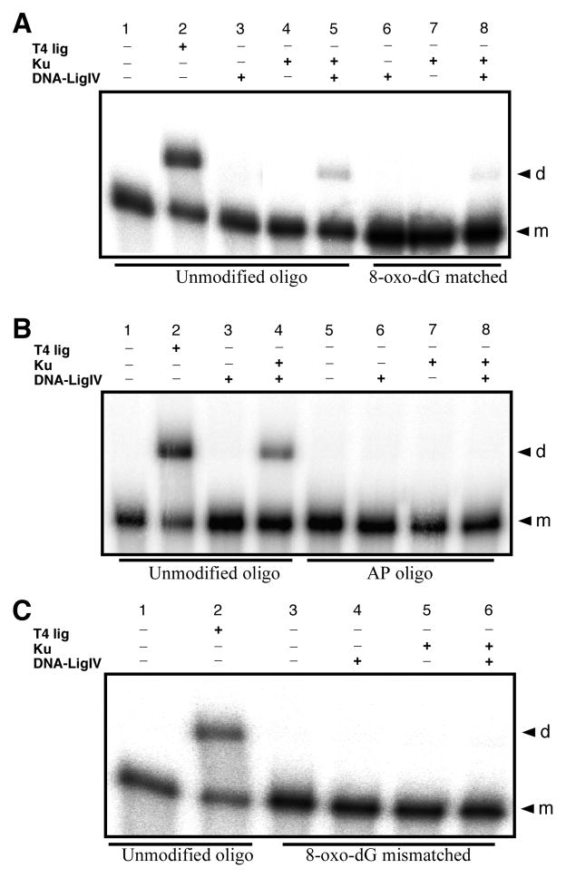
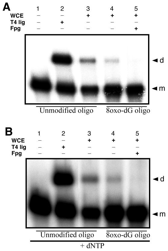
Radiation-induced DNA double-strand breaks (DSBs) are critical cytotoxic lesions that are typically repaired by nonhomologous end joining (NHEJ) in human cells. Our previous work indicated that the highly cytotoxic DSBs formed by (125)I decay possess base damage clustered within 8 to 10 bases of the break and 3'-phosphate (P) and 3'-OH ends. This study examined the effect of such structures on NHEJ in in vitro assays employing either (125)I decay-induced DSB linearized plasmid DNA or structurally defined duplex oligonucleotides. Duplex oligonucleotides that possess either a 3'-P or 3'-phosphoglycolate (PG) or a ligatable 3'-OH end with either an AP site or an 8-oxo-dG 1 nucleotide upstream (-1n) from the 3'-terminus have been examined for reparability. Moderate to severe end-joining inhibition was observed for modified DSB ends or 8-oxo-dG upstream from a 3'-OH end. In contrast, abolition of end joining was observed with duplexes possessing an AP site upstream from a ligatable 3'-OH end or for a lesion combination involving 3'-P plus an upstream 8-oxo-dG. In addition, base mismatches at the -1n position were also strong inhibitors of NHEJ in this system, suggesting that destabilization of the DSB terminus as a result of base loss or improper base pairing may play a role in the inhibitory effects of these structures. Furthermore, we provide data indicating that DSB end joining is likely to occur prior to removal or repair of base lesions proximal to the DSB terminus. Our results show that base damage or base loss near a DSB end may be a severe block to NHEJ and that complex combinations of lesions presented in the context of a DSB may be more inhibitory than the individual lesions alone. In contrast, blocked DSB 3'-ends alone are only modestly inhibitory to NHEJ. Finally, DNA ligase activity is implicated as being responsible for these effects.


Figures

Figure 1
Figure 1
HeLa extract mediated end joining of 125I decay-induced DSB terminated linear plasmid DNA. A) Typical 1% agarose gel results for HeLa cell extract mediated end joining of linear plasmid DNA. All reactions were run in duplicate. The upper panel shows end-joining results for damaged linear plasmid DNA irradiated (+) 2 M DMSO, while the lower panel presents results for plasmid linearized by irradiation (−) DMSO. The contents of the lanes in both gels are as follows: lanes 1, cell extract end-joining activity control using StuI cut plasmid DNA (blunt end); lanes 2–7 present results for the end-joining reactions using the 125I-linearized damaged plasmids as substrates; lanes 2, damaged DNA + heat inactivated WCE; lanes 3, damaged DNA + WCE; lanes 4, T4 PNK 3′-phosphatase pretreated damaged DNA alone; lanes 5, T4 PNK 3′ phosphatase pretreated damaged DNA + WCE; lanes 6, damaged DNA + T4 DNA ligase; lanes 7, damaged DNA alone. The bands on the gels are identified as follows: L, linear plasmid; C, Open circular; D, dimer; T, trimer. B) Densitometric analysis of the data presented in panel A. The column numbering of the graph corresponds to the lane numbering described for panel A. Gray bars indicate DNA irradiated (+) DMSO, black bars indicate DNA irradiated (−) DMSO. The mean of the replicates is plotted and the error bars indicate the standard deviation.
Figure 2
Figure 2
Effect of reaction-component supplementation on end joining. A) The effect of 250 μM dNTP supplementation on HeLa WCE mediated end-joining reactions conducted with either the undamaged StuI-cut positive-control linear plasmid (blunt end), or damaged linear-plasmid DNA from the (+) DMSO irradiated sample. B) HeLa WCE mediated end-joining reactions supplemented with 10 units of recombinant human DNA ligase IV/XRCC4 complex alone, or the DNA ligase IV/XRCC4 complex and 250 μM dNTPs. As in panel A, reactions were conducted with the linear StuI-cut positive-control plasmid, or the (+) DMSO irradiated damaged linear-plasmid substrates. Reactions were run in duplicate and the mean of the replicates is plotted. The error bars indicate standard deviation.
Figure 3
Figure 3
Effects of 3′-end group structure on HeLa WCE mediated end joining. Duplex oligonucleotide substrates with defined end group chemistry were formed by individually annealing oligos ag to complementary 5′-32P labeled oligo h or i (Table 1), or in one case to the 3′-32P labeled (see Materials and Methods) oligo h*. All end joining reactions were run with 15 μg WCE or 0.2 unit T4 DNA ligase as indicated. Reaction products were resolved and identified by 8% denaturing PAGE and phosphorimaging, and gels representative of 3 experiments are shown. A) Typical end joining results with the unmodified a/h control oligo duplex and the 3′-P b/h and 3′-PG c/h terminated oligo duplexes. Lane 1, negative control; lane 2, T4 DNA ligase positive control; lanes 3–5 WCE mediated reactions. B) Typical end joining results for the –1n 8-oxo-dG e/i oligo duplex. C) Typical end joining results for the –1n AP site f/h oligo duplex. D) End joining reactions complementary to those depicted in panel C, only employing the 3′-32P end labeled lower-strand in the a/h* control, and f/h* –1n AP site modified, oligo duplex substrates to assess for potential 5′-32P label loss in the previous experiments. E) Typical end joining results for the multiply damaged g/i duplex oligo structure containing a –1n 8-oxo-dG in combination with a 3′-P blocked upper-strand 3′-end. F) Typical end joining reaction results for assays using the mismatch oligo substrates exhibiting either a –1n A:C mismatch (duplex a/i), or a –1n mismatch involving the 8-oxo-dG base lesion opposite thymine (duplex e/h). G) Typical end joining results for the –1n 8-oxo-dG e/i oligo duplex (G*) matched with the d/i -1n G:C base sequence matched control (Ctrl) and WCEs as indicated. In all panels, the “d” to the right of the gel indicates dimer end joining products and the “m” indicates monomer substrate oligonucleotide.
Figure 4
Figure 4
Electrophoretic mobility shift assays (EMSA) and Ku80 immunodepletion effects. EMSAs were conducted as described in the Materials and Methods with the HeLa cell extract and the 5′-32P end labeled undamaged a/h oligo duplex (A), the 8-oxo-dG modified e/i oligo duplex (B), or the AP site modified f/h oligo duplex (C) as the protein-binding targets. The protein-bound duplex oligonucleotides were resolved in a 6% non-denaturing PAGE gel and data was obtained by phosphorimaging. Panel A is a compilation of data from two separate experiments as indicated by the vertical dashed line. In panels A – C, the letters to the right of the gel images represent the following: “a” identifies the position of the anti-Ku70 antibody mediated supershift; bands migrating between a and b in the 6th & 7th lanes of panel A and 4th & 5th lanes of panels B and C are the anti-DNA-Pkcs and anti-DNA ligase IV supershift bands as indicated. The “c” indicates the position of the HeLa extract mediated band shift, and “d” indicates the position of the unshifted duplex oligo targets. The functional effects of Ku80 immunodepletion on HeLa WCE end joining was assessed as indicated in panel D using either the unmodified a/h oligo duplex or the 3′-P terminated b/h duplex as the end joining substrates. In this case, the “d” to the right of the gel indicates the end joined dimer product and the “m” indicates the monomer substrate duplex.
Figure 5
Figure 5
End joining of modified and unmodified duplex oligonucleotide substrates with purified recombinant human Ku70/80 heterodimer and/or DNA ligase IV/XRCC4 tetrameric protein complexes. A) Comparison of end joining for the unmodified a/h control oligo duplex with either T4 DNA ligase (as an end joining positive control (0.2 U)) or Ku 70/80 (1.5 U) + DNA ligase IV/XRCC4 (1.5 U), and the equivalent reactions using the e/i –1n 8-oxo-dG oligo duplex substrate. B) Comparison of end joining for the unmodified a/h control oligo duplex with either T4 DNA ligase (0.2 U) or Ku 70/80 (1.5 U) + DNA ligase IV/XRCC4 (1.5 U), and the equivalent reactions using the f/h –1n AP site modified oligo duplex. C) Attempted end joining of the e/h –1n 8-oxo-dG:T mismatch containing oligo duplex with Ku 70/80 (1.5 U) + DNA ligase IV/XRCC4 (1.5 U). In all cases, the “d” to the right of the gel indicates the end joined dimer product and the “m” indicates the monomer substrate duplex.
Figure 6
Figure 6
Temporal sequence of DNA repair reactions at complex DNA DSB ends. HeLa WCE mediated NHEJ reactions were performed using either the unmodified a/h oligo duplex as a positive control for end joining, or the –1n 8-oxo-dG modified e/i oligo duplex. After stopping the standard reaction, the end-joined dimer products of the assay were treated with Fpg (1U, 37°C for 30 min) to test for the continued presence of 8-oxo-dG. A) Fpg treatment of the e/i oligo duplex dimer end-joining products. B) End joining and Fpg treatment of the e/i oligo duplex dimer end-joining products in the presence of 250 μM dNTPs.

References

    1. Ward JF. Some biochemical consequences of the spatial distribution of ionizing radiation-produced free radicals. Radiat Res. 1981;86:185–195. - PubMed
    1. Iliakis G. The role of DNA double strand breaks in ionizing radiation-induced killing of eukaryotic cells. Bioessays. 1991;13:641–648. - PubMed
    1. Khanna KK, Jackson SP. DNA double-strand breaks: signaling, repair and the cancer connection. Nat Genet. 2001;27:247–254. - PubMed
    1. Pastwa E, Neumann RD, Mezhevaya K, Winters TA. Repair of radiation-induced DNA double-strand breaks is dependent upon radiation quality and the structural complexity of double-strand breaks. Radiat Res. 2003;159:251–261. - PubMed
    1. Radford IR. DNA lesion complexity and induction of apoptosis by ionizing radiation. Int J Radiat Biol. 2002;78:457–466. - PubMed

Publication types