. 2011 May;17(5):537-48.
doi: 10.1089/ten.TEC.2010.0609. Epub 2011 Feb 14.

A new isolation method of human limbal progenitor cells by maintaining close association with their niche cells

Affiliations

A new isolation method of human limbal progenitor cells by maintaining close association with their niche cells

Szu-Yu Chen et al. Tissue Eng Part C Methods. 2011 May.

Abstract

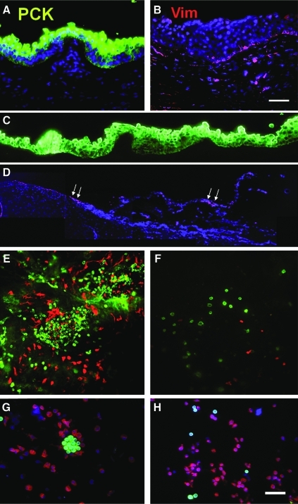
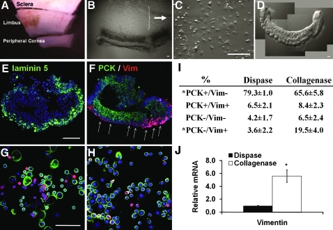
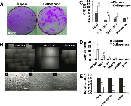
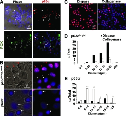
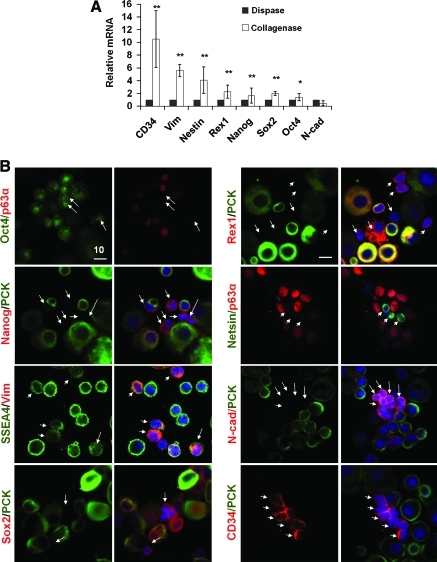
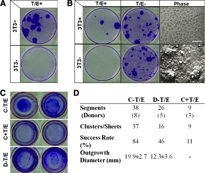
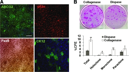
In human corneal epithelium, self-renewal and fate decision of stem cells are highly regulated in a niche microenvironment called palisades of Vogt in the limbus. Herein, we discovered that digestion with dispase, which cleaves off the basement membrane, did not remove the entire basal epithelial progenitor cells. In contrast, digestion with collagenase isolated on cluster consisting of not only entire epithelial progenitor cells but also their closely associated mesenchymal cells because of better preservation of some basement membrane matrix. Collagenase isolated more basal epithelial progenitor cells, which were p63α+ and small in the size (8 μm in diameter), and generated significantly more holoclones and meroclones on 3T3 fibroblast feeder layers than dispase. Further, collagenase isolated more small pan-cytokeratin-/p63α-/vimentin+ cells with the size as small as 5 μm in diameter and heterogeneously expressing vimentin, Oct4, Sox2, Nanog, Rex1, Nestin, N-cadherin, SSEA4, and CD34. Maintenance of close association between them led to clonal growth in a serum-free, low-calcium medium, whereas disruption of such association by trypsin/EDTA resulted in no clonal growth unless cocultured with 3T3 fibroblast feeder layers. Similarly, on epithelially denuded amniotic membrane, maintenance of such association led to consistent and robust epithelial outgrowth, which was also abolished by trypsin/EDTA. Epithelial outgrowth generated by collagenase-isolated clusters was significantly larger in diameter and its single cells yielded more holoclones on 3T3 fibroblast feeder layers than that from dispase-isolated sheets. This new isolation method can be used for exploring how limbal epithelial stem cells are regulated by their native niche cells.


Figures

FIG. 1.
FIG. 1.
Double immunofluorescence staining of pan-cytokeratins (PCK, green) and Vimentin (Vim, red). PCK+ full-thickness epithelium was present in the cryosectioned human limbal tissue (A), whereas scattered Vim+ cells were subjacent to the epithelium (B). Dispase digestion at 4°C/16 h removed the entire PCK+ epithelial sheet (C). There were no PCK+ cells in the cross section of the remaining stroma, whereas some Vim+ cells were present in the basement membrane area (marked by white arrows) and the stroma (D). Wholemount preparation of the remaining limbal stroma showed in the en face optical image aggregations of numerous PCK+ and Vim+ cells at limbus region (E), but few scattered PCK+ and Vim+ cells in the peripheral cornea (F). Cytospin preparation of cells released from the remaining stroma by collagenase digestion revealed many PCK+ clusters (G) that were associated with Vim+ cells. When these cells were further treated with T/E, PCK+ clusters disappeared and single PCK+ cells were intermixed with Vim+ cells (H). Nuclear counterstaining was performed using Hoechst 33342. Scale bar = 100 μm. T/E, trypsin/EDTA. Color images available online at www.liebertonline.com/tec
FIG. 2.
FIG. 2.
Limbal clusters obtained by collagenase digestion. A corneoscleral rim was subdivided into 12 segments, among which each was further trimmed off 1 mm above and below the limbal region (A). Each limbal segment was subjected to collagenase digestion in SHEM at 37°C for 18 h to yield a compact pigmented limbal cluster (B). After additional 24 h culturing on plastic in SHEM, the remaining cells adhered on plastic (C). Each cluster could be manually transferred by a pipette (D). Tangential sectioning of a cluster showed the preservation of laminin 5 (green) on the basal layers (E). Double immunostaining revealed that the cluster contained PCK+ cells (green) and Vim+ (red) cells, which were preferentially distributed on the basement membrane side (F, white arrows). Double immunostaining of cytospin preparations derived from collagenase-isolated clusters revealed more Vim+ cells (G, n = 1200) than those derived from dispase-isolated sheets, which had more PCK+ cells (H, n = 834). Such a difference was confirmed by counting of PCK+ cells and Vim+ cells using Zeiss image analysis (I, from 3 different bright fields and each derived from 3 donors, *p < 0.05). qRT-PCR confirmed a significant sixfold high expression of Vim mRNA in collagenase-isolated clusters than dispase-isolated sheets (J, *p < 0.05, triplicate from 3 donors). Scale bars = 100 μm. SHEM, supplemented hormonal epithelial medium; qRT-PCR, quantitative real-time polymerase chain reaction. Color images available online at www.liebertonline.com/tec
FIG. 3.
FIG. 3.
Comparison of clonal growth between dispase-isolated sheets and collagenase-isolated clusters. Significantly more rhodamine B-stained clones were generated by collagenase-isolated clusters than dispase-isolated sheets when 500 single cells were seeded in a 100-mm dish containing mitomycin C-treated 3T3 fibroblast feeder layers and cultured in SHEM for 9 days (A). Three different types of clones, that is, holoclone, meroclone, and paraclone, were identified based on the smoothness of the border and the cell size in the center of the clone with uniformly small cells, mixture of small and large cells, and uniformly large cells, respectively (see 1, 2, and 3 derived from respective insets) (B). Collagenase-isolated clusters had a significantly higher percentage of holoclones and meroclone (C, n = 3, *p < 0.05, and **p < 0.001). qRT-PCR revealed that collagenase-isolated clusters had a significant higher expression of ABCG2, Msi-1, Bmi-1, and C/EBPδ transcripts (D), but a lower transcript expression of ΔNp63α, cytokeratin 12 (CK12), connexin 43, and Pax6 (E) when compared to dispase-isolated sheets (n = 3, **p < 0.001). No significant difference was found in transcript expression of CK15 (D). Scale bar = 100 μm. Color images available online at www.liebertonline.com/tec
FIG. 4.
FIG. 4.
Correlation between cell size and p63α expression. Cytospin preparations were used to measure the cell size in diameter (μm) and for immunostaining. Double immunostaining confirmed coexpression of p63α (red) and PCK (green) in all small and medium epithelial cells (nuclear counterstaining by Hoechst 33342) and revealed the presence of small PCK−/p63α− cells (circled by white dotted lines) (A). p63α nuclear staining could be subdivided into bright (red arrows), weak (white arrows), and negative, of which the latter tended to be small (B, white arrows in the lower panel). Dispase isolated more p63αbright cells in the size from 10 to 25 μm (C, D), whereas collagenase isolated more p63α− cells in the size spreading from 8 to >25 μm (C, E). Total cells counted in Dispase n = 675 and in Collagenase n = 952, *p < 0.05, **p < 0.001). Scale bar = 25 μm in (A) and (C), and 10 μm in (B). Color images available online at www.liebertonline.com/tec
FIG. 5.
FIG. 5.
Phenotypic characterization of putative niche cells from collagenase-isolated clusters. qRT-PCR showed that collagenase-isolated clusters expressed significantly more Vim, Nestin, Rex1, Nanog, and Sox2 transcripts than dispase-isolated sheets (A, n = 3, *p < 0.05, **p < 0.001). Double immunostaining between PCK (green) or p63α (red), of which both were colocalized in epithelial cells (Fig. 4A) or between Vim (red) and other markers revealed that small nonepithelial cells shown in Figure 4 were N-cad+ (red), Sox2+ (red), Nestin+ (green), Oct4+ (green), Rex1+ (red), Nanog+ (red), CD34+ (red), and SSEA4 (green) (B, arrows). Scale bar = 10 μm. Color images available online at www.liebertonline.com/tec
FIG. 6.
FIG. 6.
Loss of clonal growth and outgrowth by disrupting close contact with niche cells by T/E. In defined keratinocyte serum-free medium, single cells derived from dispase-isolated sheets (37°C/2 h) by T/E did not generate any clonal growth as judged by crystal violet staining on day 10 in six-well plates, unless 3T3 fibroblast feeder layers were added (A). Similarly, single cells derived from the remaining stroma released by collagenase and treated with T/E (Fig. 1) also did not generate any clonal growth unless 3T3 fibroblast feeder layers were added (B, T/E+). In contrast, cells released from the remaining stroma by collagenase without T/E, which showed epithelial clusters associated with nonepithelial cells (Fig. 1), could generate vivid clonal growth without 3T3 fibroblast feeder layers (B, T/E−). Clones generated on 3T3 fibroblast feeder layers consisted of a monolayer of epithelial cells, whereas those without 3T3 feeder layers exhibited compacted small cells with scattered mounts (B, Phase). In SHEM, an outgrowth on dAM inserts in six-well plates, judged by crystal violet staining on day 7, was consistently generated by collagenase-isolated clusters without T/E (C-T/E) but abolished with T/E (C+T/E), and to a lesser extent by dispase-isolated sheets without T/E (D-T/E) (C, two representative segments from the same donor). Using more limbal segments from different donors, C-T/E gave an overall success rate of 84% and achieved a round shape outgrowth within the diameter of 19.9 ± 2.7 mm in 7 days. In contrast, C+T/E gave a success rate of 11% and no measurable diameter, whereas D-T/E yielded a 46% success rate and generated an uneven round shape with a smaller diameter of 12.3 ± 3.6 mm (D, p < 0.05). Scale bar = 100 μm in (B). dAM, denuded amniotic membrane. Color images available online at www.liebertonline.com/tec
FIG. 7.
FIG. 7.
Comparison of outgrowth on dAM generated by collagenase-isolated clusters and dispase-isolated sheets. Immunostaining confirmed universal expression of ABCG2 in cell membranes and p63α and Pax6 expression in nuclei of small basal epithelial cells, whereas CK12 was expressed by few large suprabasal cells (A). Single cells derived from the outgrowth isolated from dAM by dispase followed by T/E gave significantly more clones comprising all three classes of holoclones, meroclones, and paraclones from collagenase-isolated clusters when compared to dispase-isolated sheets (B, rhodamin B staining on day 9, *p < 0.05, **p < 0.001). Scale bar = 100 μm. Color images available online at www.liebertonline.com/tec

References

    1. Lavker R.M. Tseng S.C. Sun T.T. Corneal epithelial stem cells at the limbus: looking at some old problems from a new angle. Exp Eye Res. 2004;78:433. - PubMed
    1. Schermer A. Galvin S. Sun T.-T. Differentiation-related expression of a major 64K corneal keratin in vivo and in culture suggests limbal location of corneal epithelial stem cells. J Cell Biol. 1986;103:49. - PMC - PubMed
    1. Chen W.Y. Mui M.M. Kao W.W. Liu C.Y. Tseng S.C. Conjunctival epithelial cells do not transdifferentiate in organotypic cultures: expression of K12 keratin is restricted to corneal epithelium. Curr Eye Res. 1994;13:765. - PubMed
    1. Liu C.-Y. Zhu G. Converse R. Kao C.W.-C. Nakamura H. Tseng S.C.G. Mui M.-M. Seyer J. Justice M.J. Stech M.E., et al. Characterization and chromosomal localization of the cornea-specific murine keratin gene Krt1.12. J Biol Chem. 1994;269:24627. - PubMed
    1. Matic M. Petrov I.N. Rosenfeld T. Wolosin J.M. Alterations in connexin expression and cell communication in healing corneal epithelium. Invest Ophthalmol Vis Sci. 1997;38:600. - PubMed

Publication types

MeSH terms