Review
. 2010 Dec:1214:168-79.
doi: 10.1111/j.1749-6632.2010.05868.x.

Hearing loss: a common disorder caused by many rare alleles

Affiliations
Review

Hearing loss: a common disorder caused by many rare alleles

Dorith Raviv et al. Ann N Y Acad Sci. 2010 Dec.

Abstract

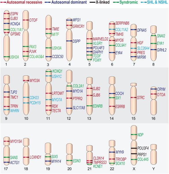
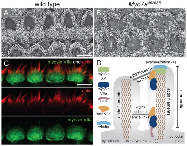
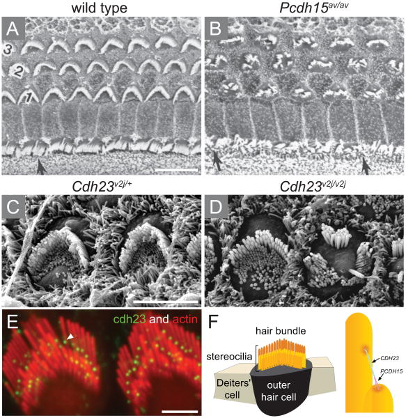
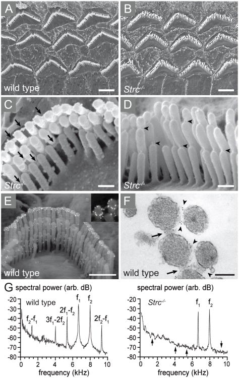
Perception of sound is a fundamental role of the auditory system. Traveling with the force of their mechanical energy, sound waves are captured by the ear and activate the sensory pathway of this complex organ. The hair cells, specialized sensory cells within the inner ear, transmit the mechanical energy into electrical nerve stimuli that reach the brain. A large number of proteins are responsible for the overarching tasks required to maintain the complex mechanism of sound sensation. Many hearing disorders are due to single gene defects inherited in a Mendelian fashion, thus enabling clinical diagnostics. However, at the same time, hearing impairment is genetically heterogeneous, with both common and rare forms occurring due to mutations in over 100 genes. The crosstalk between human and mouse genetics has enabled comprehensive studies on gene identification and protein function, taking advantage of the tools animal models have to offer. The aim of the following review is to provide background and examples of human deafness genes and the discovery of their function in the auditory system.


Figures

Figure 1
Figure 1
A scheme representing the chromosomal location of genes with mutations that cause hearing loss. The genes are classified as non-syndromic autosomal recessive (red), non-syndromic autosomal dominant (blue), X-linked genes (black), syndromic (green), and genes associated with both syndromic and non-syndromic hearing loss (light blue). Previously published in , with permission by authors.
Figure 2
Figure 2
Phenotypic effect of mutations in MYO7A. (A-B) Scanning electron micrographs of wild type (A) and (B) null allele of myosin VIIa (Myo7a4626SB) that expresses less than 1% of myosin VIIA protein, showing a near complete loss of stereocilia structure and disorganisation of the remaining hair bundle. (C) Immunolocalization of myosin VIIa is shown in the cuticular plate. (D) Schematic representation of the network of proteins that are involved in Usher syndrome within the stereocilia. Myosin VIIA is connected to the actin filaments as well as to harmonin b and to the tip links that are composed of cadherin 23 (cdh23) and protocadherin (pcdh15). Scale bars: 5 μm in (A) applies to B, 5 μm in (C). Figures (A-B) were received from Tim Self and Karen Steel and (D) was previously published in Friedman et al. 2007 with permission by authors.
Figure 3
Figure 3
The tip link of adjacent stereocilia is assembled by cadherin 23 and protocadherin. (A-D) Scanning electron micrographs of cochlear sensory epithelium of Ames waltzer (av) and waltzer (v) mice carrying mutations in Pcdh15 and Cdh23, respectively. (A) Wild type sensory epithelium is organized with one row of inner hair cell and three rows of outer hair cells with a typical ‘V’ shape arrangement of their stereocilia. (B) A spontaneous mutation of Pcdh15 leads to severely impaired organization of the hair bundles of both inner (indicated by arrows) and outer hair cells that lost the ‘V’ shape structure within their apical surface. (C) High power images of outer hair cells highlights the polarity nature of these cells characterized by hair bundles that point towards the same direction to the kinocilium. (D) The outer hair cells of Cdh23v2j/v2j mutant mice lost their planar cell polarity, showing groups of stereocilia of the surface of the cell facing different directions. Moreover, the essential staircase arrangement of the stereocilia is no longer maintained within the bundle. (E) Immunolocalization of cadherin 23 (green) is defined to the tip of the stereocilia (red) in adult guinea pig. (F) Schematic representation of an outer hair cell surrounded by two supporting cells. The hair bundle is composed of rows of stereocilia in gradual heights which create the typical staircase structure of the bundle (yellow). The tip links that connect adjacent stereocilia of lower and higher rows are composed of cadherin 23 and protocadherin 15. Ages: (A-B) P10; (C-D) P4. Scale bars: 10 μm in (A, B); 5 μm (C, D); 2 μm (E). Figures (A-B) are reprinted with permission from Alagramam et al. 2001; figures (C-D) from Di Palma et al. 2001 and figures (E-F) from Kazmierczak et al. 2007.
Figure 4
Figure 4
Stereocilin assembles the horizontal top connectors and is suggested to be essential for the nonlinearity underlying cochlear waveform distortions. (A-D) Scanning electron micrographs (SEM) of cochlear sensory outer hair cells of stereocilin deficient mice, Strc-/-. (A) Low power image of the three rows of outer hair cells in a wild type mouse. (B) The stereocilia of Strc-/- outer hair cells retain the typical ‘V’ shape but are dispersed from one another. (C) Higher magnification image of a single outer hair cell of Strc-/- mice shows that the top connectors between stereocilia (arrows) are absent in the mutant hair bundles. (D) Despite the missing top connectors, the tip links of Strc-/- hair bundles are maintained (arrow heads). (E-F) The localization of stereocilin is detected by immunogold labelling on scanning electron micrographs of a wild type outer hair cell (E), in a ring-shaped labelling around the tips of the tallest stereocilia row. Labelling can also be detected between the stereocilia within the top connectors, in a transmission electron micrograph of a stereocilin-labelled wild type outer hair cell hair bundle (F), marked with arrows. (G) Cochlear microphonic (CM) shown by frequency spectra. The black arrows within the Strc-/- mice spectra indicate the frequency points where prominent intermodulations are apparent. Ages: (A-E) P14 (F) P22. Scale bars: 2.5 μm in (A, B), 0.25 μm (C, D), 1 μm (E) and 0.2 μm in (F). Figures (A-G) are reprinted with permission from Verpy et al. 2008.
Figure 5
Figure 5
miR-96 is required for normal morphology and function of the auditory system. (A-D) Scanning electron microscopy (SEM) images of cochlear sensory hair cells of diminuendo mice depleted of miR-96. Wild type inner (A) and outer (C) hair cells exhibit normal morphology of the hair bundles with actin-rich stereocilia protrusions arranged in a typical staircase manner. (B and D) Created by ENU mutagenesis, a point mutation within the seed of miR-96 in Dmdo mice leads to altered morphology of the auditory hair cells. (B) The inner hair cells of the Dmdo/+ mutant mice show additional rows of stereocilia with a bulged and rounded cell surface. (D) Dmdo/+ outer hair cells lost their staircase arrangement while stereocilia of the first row of the bundle are taller than the middle rows. (E) In situ hybridization (ISH) revealed the spatial expression of miR-96 within the auditory apparatus defined to the cochlear inner and outer hair cells (dark blue). (F) Schematic illustration combining the top and cross view of the cochlear sensory epithelium, also known as the organ of Corti. The hair cells (green) are shown in their typical arrangement of one inner and three outer rows. The hair bundle of each hair cell is apparent on its surface (black). A wide array of supporting cells are essential surrounding the hair cells including Dieters' (blue) Hensens and inner sulcus cells (purple). The pillar cells (brown) create the tunnel of Corti that separate between the inner and outer rows of hair cells. Ages: (A-D) P28 (E) P0. Scale bars: 3 μm in (A-D), 5 μm in (E). Figures (A-E) are reprinted with permission from Lewis et al. 2009 and figure (F) was previously published in Dror and Avraham 2009.

References

    1. Petit C, Richardson GP. Linking genes underlying deafness to hair-bundle development and function. Nat Neurosci. 2009;12:703–710. - PMC - PubMed
    1. Petit C, Levilliers J, Hardelin JP. Molecular genetics of hearing loss. Annu Rev Genet. 2001;35:589–646. - PubMed
    1. Gorlin RJ. Genetic Hearing Loss-A Brief History. In: Gorlin RJ, Toriello HV, Cohen MM, editors. Hereditary Hearing Loss and its Syndromes. Oxford University Press; New York: 1995. pp. 3–4.
    1. Cranefield PF, Federn W. Paulus Zacchias on mental deficiency and on deafness. Bull N Y Acad Med. 1970;46:3–21. - PMC - PubMed
    1. Dow GS, Poynter CI. The Dar family. Eugen News. 1930;15:128–130.

Publication types