. 2010 Dec 22:8:152.
doi: 10.1186/1741-7007-8-152.

Drosophila EGFR pathway coordinates stem cell proliferation and gut remodeling following infection

Affiliations

Drosophila EGFR pathway coordinates stem cell proliferation and gut remodeling following infection

Nicolas Buchon et al. BMC Biol. .

Abstract

Background: Gut homeostasis is central to whole organism health, and its disruption is associated with a broad range of pathologies. Following damage, complex physiological events are required in the gut to maintain proper homeostasis. Previously, we demonstrated that ingestion of a nonlethal pathogen, Erwinia carotovora carotovora 15, induces a massive increase in stem cell proliferation in the gut of Drosophila. However, the precise cellular events that occur following infection have not been quantitatively described, nor do we understand the interaction between multiple pathways that have been implicated in epithelium renewal.

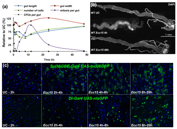
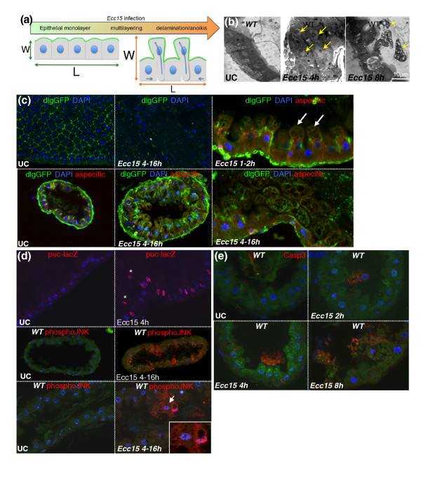
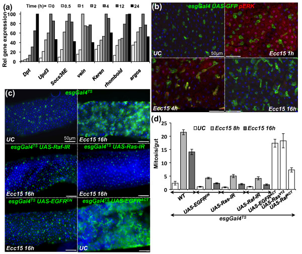
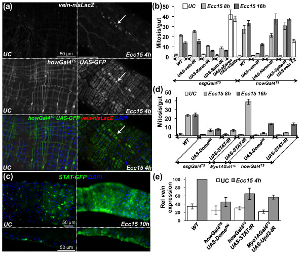
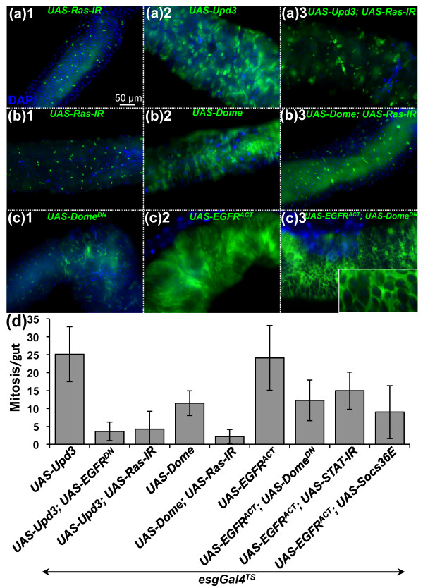
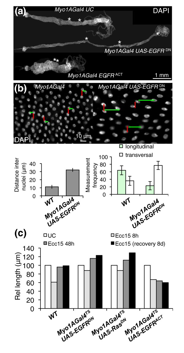
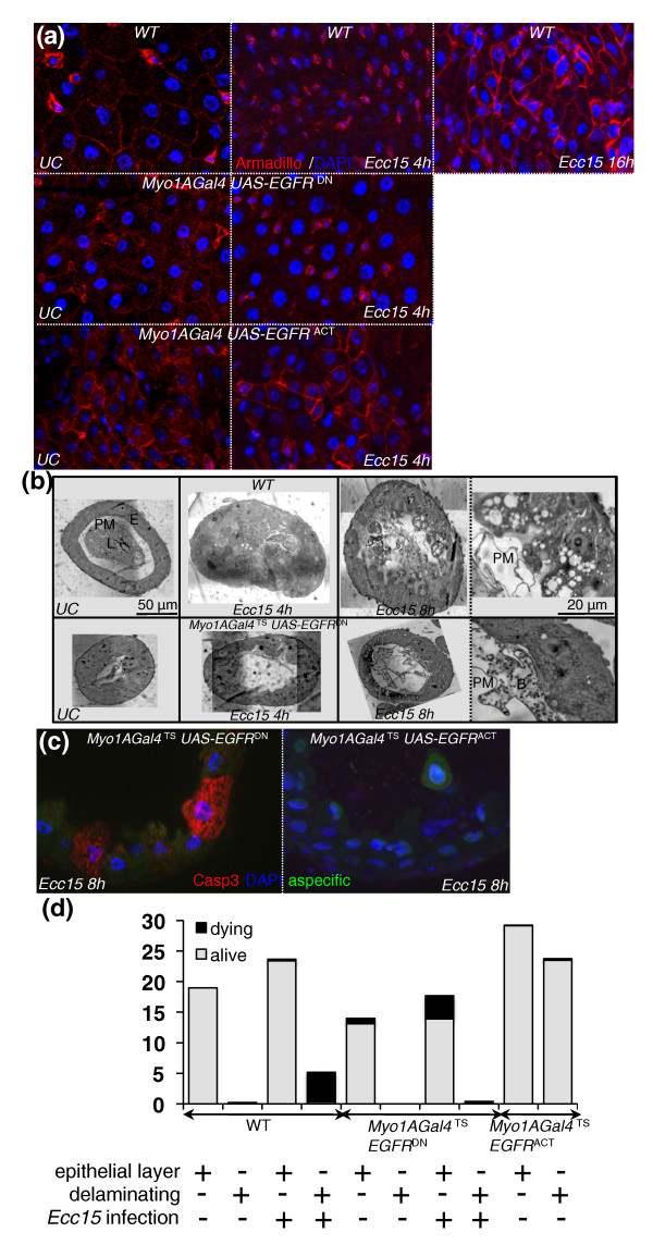
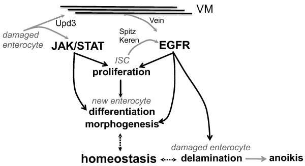
Results: To understand the process of infection and epithelium renewal in more detail, we performed a quantitative analysis of several cellular and morphological characteristics of the gut. We observed that the gut of adult Drosophila undergoes a dynamic remodeling in response to bacterial infection. This remodeling coordinates the synthesis of new enterocytes, their proper morphogenesis and the elimination of damaged cells through delamination and anoikis. We demonstrate that one signaling pathway, the epidermal growth factor receptor (EGFR) pathway, is key to controlling each of these steps through distinct functions in intestinal stem cells and enterocytes. The EGFR pathway is activated by the EGF ligands, Spitz, Keren and Vein, the latter being induced in the surrounding visceral muscles in part under the control of the Janus kinase/signal transducer and activator of transcription (JAK/STAT) pathway. Additionally, the EGFR pathway synergizes with the JAK/STAT pathway in stem cells to promote their proliferation. Finally, we show that the EGFR pathway contributes to gut morphogenesis through its activity in enterocytes and is required to properly coordinate the delamination and anoikis of damaged cells. This function of the EGFR pathway in enterocytes is key to maintaining homeostasis, as flies lacking EGFR are highly susceptible to infection.

Conclusions: This study demonstrates that restoration of normal gut morphology following bacterial infection is a more complex phenomenon than previously described. Maintenance of gut homeostasis requires the coordination of stem cell proliferation and differentiation, with the incorporation and morphogenesis of new cells and the expulsion of damaged enterocytes. We show that one signaling pathway, the EGFR pathway, is central to all these stages, and its activation at multiple steps could synchronize the complex cellular events leading to gut repair and homeostasis.


Figures

Figure 1
Figure 1
Ingestion of Erwinia carotovora carotovora 15 (Ecc15) induces dramatic morphological changes to the gut of Drosophila. (a) Quantitative measurements of the gut at different time points after infection reveal that Ecc15 induces a dramatic remodeling of the gut. The midgut lengths and widths, the total number of cells and the number of mitotic stem cells (phosphohistone H3 (PH3)-positive cells) along the midgut are shown. Measurements of colony-forming units (CFUs) per gut of Ecc15 at corresponding time points are also shown. (b) Representative images of guts dissected from wild-type (WT), unchallenged (UC) or Ecc15-infected flies stained with 4',6-diamidino-2-phenylindole (DAPI) and observed by light microscopy at ×10 original magnification. Gut length is decreased at 4 hours after infection, but returns to unchallenged levels by 48 hours. MT, Malpighian tubules; HG, hindgut. (c) Expression of green fluorescent protein (GFP) under the control of enteroblast (Su(H)GBE-Gal4; UAS-mcd8GFP) or intestinal stem cell (ISC) (delta-Gal4; UAS-nlsGFP) specific reporter genes was monitored following infection with Ecc15. Soon after infection (> 2 hours), expansion of Su(H)GBE-Gal4; UAS-mcd8GFP GFP signal was observed along the gut, reflecting the rapid differentiation of enteroblasts into larger enterocytes. The expansion of delta-Gal4; UAS-nlsGFP was observed only after 4 hours, indicative of ISC proliferation.
Figure 2
Figure 2
Ingestion of Ecc15 results in the delamination of enterocytes and anoikis. (a) A scheme describing the mechanism by which delamination of enterocytes could account for the shortening and widening of the gut. W, width; L, length. (b) Infection results in an intense multilayering (arrows) and the blebbing (stars) and delamination of enterocytes following ingestion of Ecc15. Representative images of UC and Ecc15 infected (t = 4 and 8 hours) guts are shown. Histological sections of the anterior midgut region were analyzed by light microscopy at ×63 original magnification. (c) Localization of the septate junction marker Discs large (dlgGFP) reveals a regular pattern in UC WT flies. Cross section of the gut shows that Dlg is located apically between enterocytes. Infection with Ecc15 disrupts Dlg localization in the gut. Cell blebbing (white arrows) occurs apically to the Dlg compartment. (d) Immunostaining of guts expressing the c-Jun N-terminal kinase (JNK)-responsive reporter gene puc-lacZ revealed that ingestion of Ecc15 results in the activation of the JNK pathway in most enterocytes and all delaminating cells. Additionally, immunostaining of sections of guts from WT flies with antibodies directed against the phospho-form of JNK (red) reveals continued JNK activation in delaminating enterocytes (4 to 16 hours postinfection with Ecc15). Fragmented nuclei were observed in some of these JNK-positive cells (inset). (e) Immunostaining of histological sections of WT flies with antibodies directed against the cleaved form of caspase 3 shows that cells undergo apoptosis only after they have detached from the epithelium. This signal was detected during the duration of the infectious process in cells at varying stages of delamination and anoikis.
Figure 3
Figure 3
The epidermal growth factor receptor (EGFR) pathway is induced in the gut and is required for ISC proliferation triggered by infection. (a) Reverse transcriptase-quantitative polymerase chain reaction (RT-qPCR) analysis of gut extracts shows that genes encoding components of the Imd (Diptericin (Dpt)), Janus kinase/signal transducer and activator of transcription (JAK/STAT) (upd3, Socs36E) and EGFR (vein, Keren, rhomboid, argos) pathways are induced upon oral ingestion with Ecc15. Values were normalized to RpL32 and set relative to their own maximum induction levels. (b) Immunostaining of guts of esgGal4 UAS-GFP flies with antibodies directed against the phospho-form of the extracellular signal-regulated kinase (ERK) kinase (red) and GFP (green) reveals that the ERK kinase was activated in enterocytes 1 hour postinfection and in both progenitor cells (green) and enterocytes at 4 hours. At 16 hours, no phospho-ERK signal was detected. (c) Ingestion of Ecc15 induces a marked increase in the number of esgGal4TS, UAS-GFP-positive cells (indicative of epithelium renewal), which was not observed when double-stranded RNA (dsRNA or RNAi) or dominant-negative forms of members of the EGFR pathway (UAS-EGFRDN, UAS-Ras-IR or UAS-Raf-IR) were expressed in ISCs. Overexpression of an active form of EGFR (UAS-EGFRACT) in ISCs is sufficient to induce a high level of epithelium renewal in the absence of infection. (d) Quantification of PH3-positive cells per midgut shows an increase in the number of mitotic cells upon Ecc15 infection in WT flies, but not in flies with reduced EGFR activity in ISCs (UAS-EGFRDN, UAS-Ras-IR or UAS-Raf-IR; P < 0.05). Overexpression of a constitutively activated form of EGFR in ISCs increases the mitotic index (esgGal4TS, UAS-EGFRACT, UAS-RasV12 or UAS-RafACT; P < 0.05). Mean values of five experiments (N = 10 to 20 guts each) ± SE are shown. Analysis of variance (ANOVA) F = 58.64. df = 14. P < 0.0001.
Figure 4
Figure 4
The JAK/STAT pathway is required for vein expression in the visceral muscles upon infection with Ecc15. (a) Immunostaining against lacZ and GFP of guts derived from vein-lacZ; howGal4TS UAS-GFP flies reveals than vein (nuclear signal) is induced upon infection in the circular visceral muscles. (b) RNAi silencing of vein in visceral muscles and reduction of Keren and Spitz in precursor cells blocked infection-induced proliferation (P < 0.05). Conversely, ectopic expression of the three EGFs (UAS-sKeren, UAS-sSpitz and UAS-vein1.2) was sufficient to trigger proliferation (P < 0.05). Mean values from five experiments (N = 10-20 guts each) ± SE are shown. ANOVA F = 62.96. df = 26. P < 0.0001. (c) Infection induced the JAK/STAT reporter gene, STAT-GFP, in visceral muscles in addition to ISCs. Representative images of UC and Ecc15-infected guts are shown. Guts of STAT-GFP flies were stained with DAPI and examined by fluorescence microscopy at ×20 original magnification. The microscopic focus was set to the external layer of the gut (top). Transverse sections show that expression of STAT-GFP is localized to the circular visceral muscle and progenitor cells (bottom). (d) The JAK/STAT pathway is required in both ISCs and visceral muscles to promote ISC proliferation. RNAi directed against STAT92E or expression of a dominant-negative form of Domeless in ISCs (esgGal4TS) or muscles (howGal4TS), but not enterocytes (Myo1AGal4TS) strongly reduced the number of mitotic ISCs in the guts of flies infected with Ecc15 (t = 16 hours; P < 0.05). Mean values from five experiments (N = 10-20 guts each) ± SE are shown. ANOVA F = 50.74. df = 15. P < 0.0001. (e) The induction of vein upon Ecc15 infection was reduced in flies with reduced JAK/STAT signaling in visceral muscles (P < 0.05). Tissue-specific silencing of upd3 in enterocytes also reduced vein expression (P < 0.05). RT-qPCR analysis of gut extracts from UC and Ecc15-infected flies (t = 8 hours). Levels of vein expression were normalized to RpL32. Mean values from four experiments (N = 20 guts each) ± SE are shown. ANOVA F = 14.83. df = 7. P < 0.0001.
Figure 5
Figure 5
The EGFR and JAK/STAT pathways synergize to promote ISC proliferation. Expression of the esgGal4TS, UAS-GFP reporter gene (a-c) and quantification of PH3-positive cells per midgut (d) were monitored in UC flies. Overexpression of upd3 (A2) or domeless (B2) in ISCs induced higher levels of epithelium renewal in UC flies, which was reduced by depletion of EGFR or Ras activity (A3 and B3). Conversely, the high levels of epithelium renewal induced by overexpressing an activated form of the EGFR (C2) in ISCs was slightly reduced by coexpression of a negative regulator of the JAK/STAT pathway (UAS-Socs36E), a dominant-negative form of Domeless (UAS-DomeDN) or a UAS-STAT-IR construct (C3 and D). Ectopic expression of the EGFR in ISCs expressing a dominant-negative form of Domeless did not rescue the differentiation defect caused by the lack of JAK/STAT activity, but instead aggravated the expansion of undifferentiated escargot-GFP precursor cells (C3 and inset).
Figure 6
Figure 6
The EGFR pathway is required in enterocytes for proper gut morphogenesis. (a) Midguts with enterocytes depleted of the EGFR pathway (Myo1AGal4, UAS-EGFRDN) are longer and thinner than WT guts. Conversely, guts from flies expressing a constitutive form of EGFR are shorter and wider. The effect of EGFR is most pronounced in the copper cell region (borders of which are indicated with asterisks). Representative images were taken of dissected guts from 3-day-old to 4-day-old flies stained with DAPI. Guts from flies expressing a constitutive form of EGFR are approximately half the length of WT UC flies. (b) Nuclear staining of guts with EGFR-deficient enterocytes reveals a decrease in cell density and flattening of nuclei as compared to WT guts. Quantification of the mean distance between the nearest adjacent nuclei in WT and EGFR-depleted guts. Measures were taken in the region around the copper cells (middle midgut), where the effect of EGFR is most pronounced (Additional file 11C). Additionally, the orientation of the distance vector between the two nearest nuclei switches from longitudinal to transversal, indicating a change in epithelial geometry (see model, Additional file 11D). (c) Relative length of WT guts, guts with enterocytes depleted for the EGFR pathway or guts with activated EGFR in enterocytes. Constructs were placed under the control of a thermosensitive enterocyte driver (Myo1AGal4TS). Adult flies were switched from 18°C (Gal4 nonfunctional) to 29°C (Gal4 functional) 3 days before infection or before initial measurements were taken. Guts depleted for the EGFR pathway (UAS-EGFRDN or UAS-RasDN) in enterocytes shrink less upon infection with Ecc15. After a recovery phase of 2 or 8 days, the guts are 25% longer than their WT counterparts. Conversely, guts of flies with ectopic activation of EGFR (UAS-EGFRACT) in enterocytes do not elongate in response to infection and are 40% shorter than WT guts.
Figure 7
Figure 7
The EGFR pathway is required for enterocyte delamination upon infection. (a) Immunostaining of gut epithelium with Armadillo (red) and DAPI (blue). In WT flies, the Armadillo signal was strong in progenitors and lower in enterocytes. Upon infection, Armadillo staining disappeared from enterocyte membranes, but remained intense in progenitor cells. At later stages, both progenitors and newly synthesized enterocytes displayed a strong Armadillo signal. Guts with enterocytes depleted of EGFR activity (UAS-EGFRDN) displayed low Armadillo staining in enterocytes that did not change upon infection. In the absence of infection, guts with activated EGFR (UAS-EGFRACT) in enterocytes exhibited a pattern of Armadillo staining similar to Ecc15-infected WT guts. (b) Histological sections of guts from flies with WT (top) and EGFR-depleted enterocytes (bottom) with and without infection. A strong multilayering of epithelial cells (t = 4 hours), followed by their blebbing, and delamination (t = 8 hours) are observed in WT guts following infection. Delaminating cells contain multiple large vacuoles. In contrast, the multilayering, blebbing and delamination of enterocytes was not observed in guts from flies depleted of EGFR in enterocytes. PM, peritrophic matrix; L, lumen; E, enterocyte; B, bacteria. (c) In contrast to WT (Figure 2e), apoptotic enterocytes (anticleaved caspase 3-positive) were detected within the epithelial layer in flies with enterocytes depleted of EGFR activity (UAS-EGFRDN). Neither delaminated cells nor enterocytes within the epithelium were apoptotic in guts with activated EGFR (UAS-EGFRACT) in enterocytes. (d) Quantification of delaminating and apoptotic cells in the guts of flies with WT enterocytes, enterocytes depleted of EGFR activity (UAS-EGFRDN) or enterocytes expressing an activated form of EGFR (UAS-EGFRACT). Cells within the epithelium layer and delaminating cells were counted from three histological sections (N = 16 guts) for each genotype in UC and Ecc15-infected flies. The proportions of living and dead cells per section were determined with anticleaved caspase 3 and DAPI staining.
Figure 8
Figure 8
Regulation of epithelium renewal by the JAK/STAT and EGFR pathways. Upon infection, damaged enterocytes release Upd3. This cytokine activates the JAK/STAT pathway in both progenitor cells (ISCs) and in the surrounding visceral muscles (VMs). JAK/STAT activation in VMs participates in the induction of the EGF vein. In addition, two EGFs, Keren and Spitz, are also secreted by the epithelium. The activation of both the JAK/STAT and EGFR pathways in progenitor cells stimulates their proliferation. The JAK/STAT pathway has a unique role in enteroblast differentiation, while the EGFR pathway is required in enterocytes for proper morphogenesis and delamination of damaged enterocytes following infection with Ecc15. Other ligands of the JAK/STAT pathway (Upd1 and Upd2) may also participate in bacteria-induced ISC proliferation.

References

    1. Kim M, Ashida H, Ogawa M, Yoshikawa Y, Mimuro H, Sasakawa C. Bacterial interactions with the host epithelium. Cell Host Microbe. 2010;8:20–35. doi: 10.1016/j.chom.2010.06.006. - DOI - PubMed
    1. Turner JR. Intestinal mucosal barrier function in health and disease. Nat Rev Immunol. 2009;9:799–809. doi: 10.1038/nri2653. - DOI - PubMed
    1. Potten CS, Kellett M, Rew DA, Wilson GD. Measurement of in vivo proliferation in human colorectal mucosa using bromodeoxyuridine. Gut. 1992;33:71–78. doi: 10.1136/gut.33.1.71. - DOI - PMC - PubMed
    1. Ohlstein B, Spradling AC. The adult Drosophila posterior midgut is maintained by pluripotent stem cells. Nature. 2006;439:470–474. doi: 10.1038/nature04333. - DOI - PubMed
    1. Micchelli CA, Perrimon N. Evidence that stem cells reside in the adult Drosophila midgut epithelium. Nature. 2006;439:475–479. doi: 10.1038/nature04371. - DOI - PubMed

Publication types

MeSH terms