Review
. 2011 Apr 1;508(1):1-12.
doi: 10.1016/j.abb.2010.12.017. Epub 2010 Dec 19.

Tyrosine hydroxylase and regulation of dopamine synthesis

Affiliations
Review

Tyrosine hydroxylase and regulation of dopamine synthesis

S Colette Daubner et al. Arch Biochem Biophys. .

Abstract

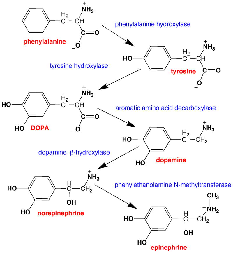
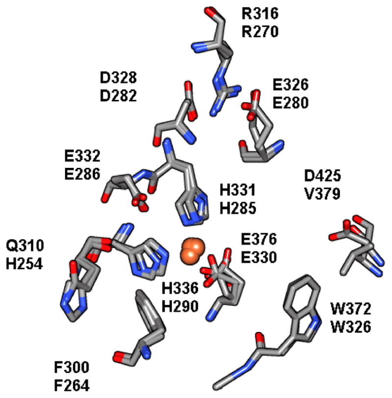
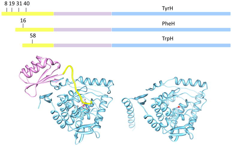
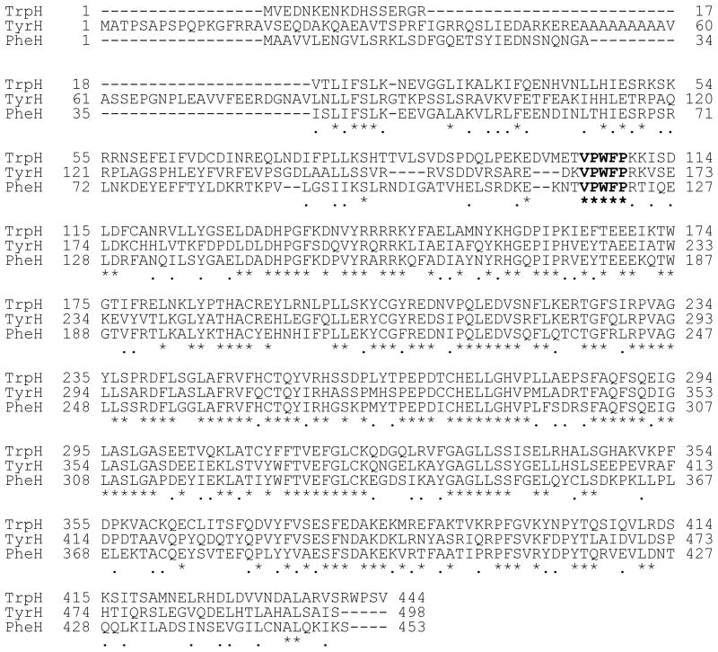
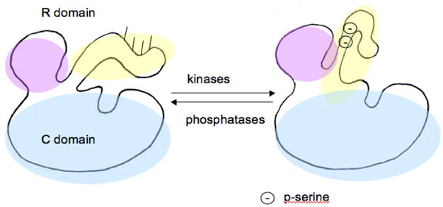
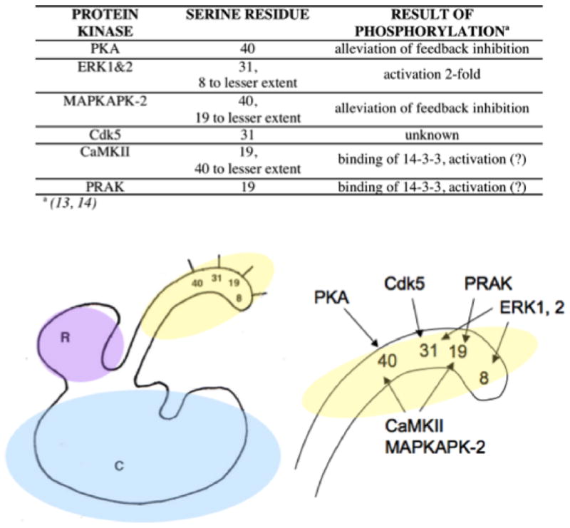
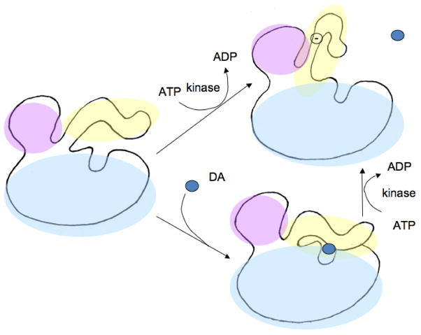
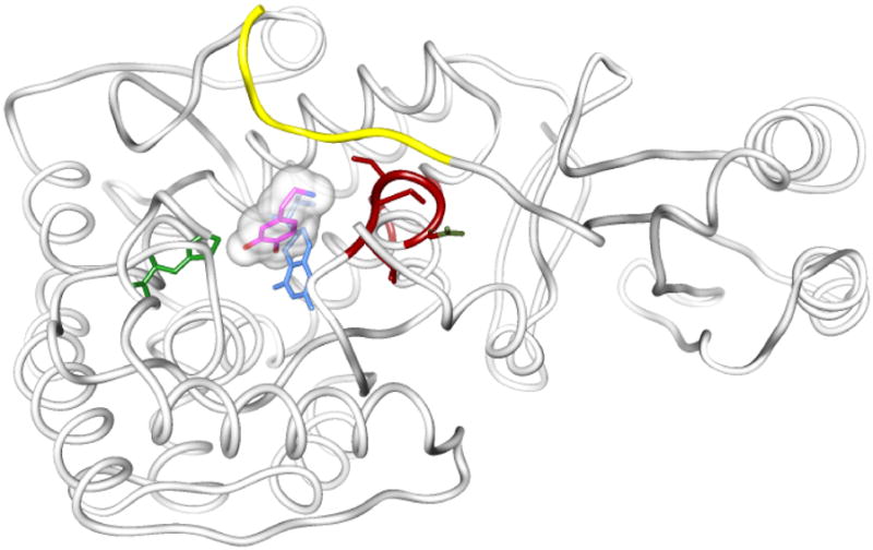
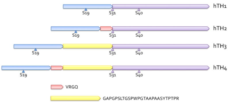
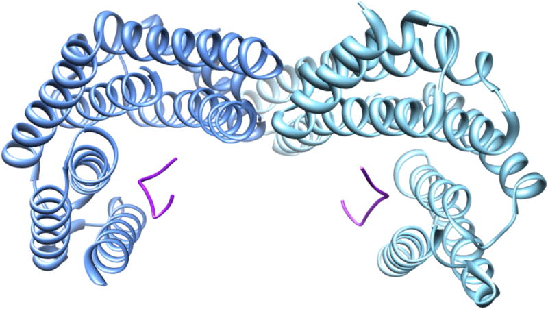
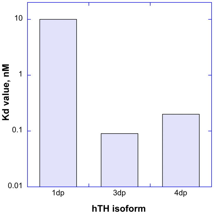
Tyrosine hydroxylase is the rate-limiting enzyme of catecholamine biosynthesis; it uses tetrahydrobiopterin and molecular oxygen to convert tyrosine to DOPA. Its amino terminal 150 amino acids comprise a domain whose structure is involved in regulating the enzyme's activity. Modes of regulation include phosphorylation by multiple kinases at four different serine residues, and dephosphorylation by two phosphatases. The enzyme is inhibited in feedback fashion by the catecholamine neurotransmitters. Dopamine binds to TyrH competitively with tetrahydrobiopterin, and interacts with the R domain. TyrH activity is modulated by protein-protein interactions with enzymes in the same pathway or the tetrahydrobiopterin pathway, structural proteins considered to be chaperones that mediate the neuron's oxidative state, and the protein that transfers dopamine into secretory vesicles. TyrH is modified in the presence of NO, resulting in nitration of tyrosine residues and the glutathionylation of cysteine residues.


Figures

Figure 1
Figure 1
The biosynthetic pathway for the catecholamine neurotransmitters. Phenylalanine hydroxylase converts phenylalanine to tyrosine, tyrosine hydroxylase hydroxylates tyrosine to L-DOPA. DOPA is converted to dopamine by aromatic amino acid decarboxylase. Dopamine-β-hydroxylase hydroxylates dopamine to norepinephrine, which is methylated to epinephrine by phenylethanolamine N-methyltransferase. Tyrosine hydroxylase is the rate-limiting enzyme of the pathway.
Figure 2
Figure 2
The active sites of PheH (1DMW) and TyrH (1TOH) are overlaid on each other and the residues that play identifiable roles in catalysis are highlighted. The top # in each pair of residues is the residue in TyrH and the bottom # is the residue in PheH. In particular, histidines 331 and 336 (285 and 290 in PheH) and glutamate 376 (330) are the ligands to the iron atom. Residues on the left in this view, gln310/his254, phe300/264, and glu332/286 form the binding site for tetrahydrobiopterin, and on the right, arg316/270, asp328/282/ and asp425/val379 form the binding site for the aromatic amino acid.
Figure 3
Figure 3
The domain structure of the aromatic amino acid hydroxylases. At top: the AAAHs consist of three domains: regulatory, catalytic, and tetramerization. The amino terminal regulatory domains differ in length (~150, TyrH; ~115, PheH; and ~105, TrpH) and contain regulatory serine residues at different positions. The portions of the R domains that are less similar among the three enzymes and that contain the regulatory serine residues are in yellow. The portions of the R domains that display some homology are in lilac. The catalytic cores are similar in length and sequence; they appear in light blue. The leucine-zipper-style tetramerization domains are at the very carboxyl termini, and are not emphasized in this figure. Left: The crystal structure of PheH (1PHZ), color coded to highlight the domain structure. The catalytic core is shown in blue, and the iron with its ligands are displayed as a sphere and stick models. The regulatory domain is above and offset to the left of the catalytic domain, and is bicolored. The R domain region that is somewhat homologous to the R domains of TyrH and TrpH is shown in lilac. Amino terminal to it, one long strand shown in yellow traverses the space to the active site and covers its opening. This region contains residues 20 to 34 of PheH. This crystal structure does not show the position of the amino terminal 19 amino acids, presumably due to flexibility. Right: The crystal structure of TyrH (1TOH). Because the R domain of TyrH has not been crystallized, presumably due to its flexibility, only the catalytic domain and the tetramerization domain are seen. The active site iron and its ligands are displayed as a sphere and stick models. This protein is missing its 155 amino terminal amino acids, therefore, its R domain is absent. The two proteins are viewed from approximately the same angle to facilitate comparison.
Figure 4
Figure 4
Clustal W alignment of the amino acid sequences of the rat aromatic amino acid hydroxylases. An asterisk below the three sequences denotes identity, and a period indicates similarity. The C domain begins near the end of the third set of rows, that is, near position 105 for TrpH, 164 for TyrH, and 117 for PheH, where the sequence VPWFP appears in bold type for all three enzymes.
Figure 5
Figure 5
A model for the mechanism of activation by phosphorylation of serine residues in the R domain of TyrH. The catalytic core is indicated in light blue. The R domain is in lavender with the mobile portion in yellow. It contains serines that become phosphorylated by kinases, at positions 8, 19, 31, and 40. Upon phosphorylation of the R domain it moves out of its position obstructing access to the active site. Phosphatases then return the R domain to its inactivating position. Only 2 serine phosphates are shown for clarity.
Figure 6
Figure 6
Simplified map of the reactivity of some protein kinases with the serine residues of the R domain of TyrH. Ser40 is modified by PKA, CaMKII, and MAPKAPK-2. Ser31 is modified by ERK1 & 2 and Cdk5. PRAK labels ser19, as do CaMKII, and MAPKAPK-2. Ser8 is modified by ERK1 but it is not certain that the reaction has an effect on TyrH activity. Extensive coverage of these phosphorylation events have been covered in previous reviews (14, 25)
Figure 7
Figure 7
On left, PheH with tetrahydrobiopterin, dopamine, and thienylalanine bound. Three structures were overlaid to create these images: 1PHZ, which contains the R domain; 4PAH, which contains catecholamine; and 1KW0, which contains tetrahydrobiopterin, and thienylalanine, a phenylalanine analog. The pterin is in blue, the dopamine is in orange, and the thienylalanine is in green. Clearly the dopamine and biopterin sites overlap, and the aromatic amino acid site is separate. At right are the same structures in a higher magnification, turned slightly to illustrate the proximity of the catecholamine to the R domain.
Figure 8
Figure 8
Drawing of three different possible regulatory domain configurations. The regulatory domain is lavender with a mobile yellow loop, and the catalytic domain is light blue. Dopamine is represented by a blue circle. Left, unphosphorylated TyrH; the R domain is very flexible. Lower right, TyrH with dopamine bound, chelated to active site iron. The R domain is in a more rigid conformation, less accessible to proteases, and obstructing entrance of substrates. Upper right, TyrHpser40 with salt bridge between some acidic residue and phosphoserine40, exposing the entrance to the active site. TyrHpser40 has released the catecholamine which was bound prior to phosphorylation, the flexible loop is in a different configuration that makes arg37 and arg38 less accessible to trypsin but exposing arg33 and arg49.
Figure 9
Figure 9
Peptides of PheH corresponding to the peptides of TyrH identified by hydrogen-deuterium exchange. The ribbon is derived from crystals of PheH missing 19 amino terminal amino acids (1PHZ). Overlaid on that structure are the structures of PheH with dopamine (5PAH), BH4 and thienylalanine (1KW0), with only the small molecules shown. Thienylalanine is in green, dopamine in magenta with its amino nitrogen in blue, and tetrahydrobiopterin is blue. The region of the R domain that is homologous to TyrH’s gly36-glu50 is colored yellow. The region of PheH that is homologous to TyrH positions 295–299 is colored red.
Figure 10
Figure 10
Drawing of the R domains of the human isoforms of TyrH illustrating their structural differences. Amino termini are to the left. hTH1 is identical in length to rat TyrH, and appears first, with the regulatory serines shown. hTH2 has four additional amino acids after met30, and those amino acids are shown as a short pink segment of the R domain. hTH3 has 27 additional amino acids, shown as a yellow segment, included after met30. hTH4 has both additional segments so has 31 amino acids more than hTH1. These additional amino acids are included via alternative mRNA splicing. Since the additional amino acids come immediately before ser31, the serine residues homologous to ser31 and ser40 have different numbers in hTH’s 2, 3, and 4, but are rarely referred to by these numbers.
Figure 11
Figure 11
Crystal structure of the dimer of 14-3-3ζ protein with phosphopeptides bound (1QJA).
Figure 12
Figure 12
Dissociation constants measured for the binding of bovine 14-3-3 ζ protein to human TyrH isoforms 1, 3, and 4 phosphorylated at ser19 and ser40.
Figure 13
Figure 13
Two views of possible orientations of 14-3-3 protein and the R domain of TyrH. The figure was composed using the structure of PheH (1PHZ) since no TyrH R domain structure is available. The green ribbons are a tetramer of TyrH (1TOH). The blue and red ribbons are 2 monomers of PheH superimposed upon the TyrH tetramer. One monomer of a 14-3-3ζ dimer (1QJA) is colored purple and the other is gold. The views represent the model at two positions rotated 90° on the x-axis.
Figure 14
Figure 14
Locations of the three tyrosine residues of TyrH that become nitrated as a result of reaction with peroxyitrate. The three tyrosine residues are at positions 423, 428, and 432 and are shown in dark blue. The active site it identifiable by the bound iron and the ligands that surround it. The figure illustrates that each tyrosine residue is very exposed to solvent.
Figure 15
Figure 15
Locations of the seven cysteine residues of TyrH. At left is the structure of TyrH overlaid on the structure of PheH; PheH is included to keep in mind where the R domain would be. The cysteine residues are shown as spheres and the sulfur atoms are yellow. At right is the same structure but TyrH is represented as a partially transparent surface rendering to illustrate the relative exposure of the cysteine residues.

Similar articles

Cited by

References

    1. Molinoff PB, Axelrod J. Biochemistry of catecholamines. Annual Review of Biochemistry. 1971;40:465–500. - PubMed
    1. Weiner N. Tyrosine-3-monooxygenase (tyrosine hydroxylase) In: Youdim MBH, editor. Aromatic amino acid hydroxylases and mental disease. John Wiley & Sons, Ltd; New York: 1979. pp. 141–190.
    1. Tripp G, Wickens JR. Neurobiology of adhd. Neuropharmacology. 2009;57:579–589. - PubMed
    1. Arnsten AFT. Catecholamine regulation of the prefrontal cortex. J Psychopharmacol. 1997;11:151–162. - PubMed
    1. Assadi SM, Yücel M, Pantelis C. Dopamine modulates neural networks involved in effort-based decision-making. Neuroscience & Biobehavioral Reviews. 2009;33:383–393. - PubMed

LinkOut - more resources